 Movie
Movie Controller


+ Open data

- Basic information
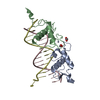
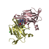
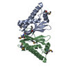
| Entry | Database: PDB / ID: 4a8x | ||||||
|---|---|---|---|---|---|---|---|
| Title | Structure of the core ASAP complex | ||||||
 Components Components |
| ||||||
 Keywords Keywords | TRANSCRIPTION / SPLICING / RNA PROCESSING / NONSENSE MEDIATED DECAY / NMD / HDAC / HISTONE DEACETYLATION | ||||||
| Function / homology |  Function and homology information Function and homology informationApoptotic cleavage of cellular proteins / ASAP complex / mRNA Splicing - Major Pathway / mRNA Splicing - Major Pathway / HDACs deacetylate histones / regulation of mRNA processing / mRNA 3'-end processing / Transport of Mature mRNA derived from an Intron-Containing Transcript / RNA Polymerase II Transcription Termination / nuclear-transcribed mRNA catabolic process, nonsense-mediated decay ...Apoptotic cleavage of cellular proteins / ASAP complex / mRNA Splicing - Major Pathway / mRNA Splicing - Major Pathway / HDACs deacetylate histones / regulation of mRNA processing / mRNA 3'-end processing / Transport of Mature mRNA derived from an Intron-Containing Transcript / RNA Polymerase II Transcription Termination / nuclear-transcribed mRNA catabolic process, nonsense-mediated decay / precatalytic spliceosome / regulation of alternative mRNA splicing, via spliceosome / histone deacetylase complex / negative regulation of mRNA splicing, via spliceosome / Nonsense Mediated Decay (NMD) enhanced by the Exon Junction Complex (EJC) / catalytic step 2 spliceosome / mRNA Splicing - Major Pathway / RNA splicing / regulation of autophagy / mRNA 3'-UTR binding / mRNA splicing, via spliceosome / Regulation of expression of SLITs and ROBOs / mRNA processing / transcription corepressor activity / transcription regulator complex / nucleic acid binding / nuclear speck / nuclear body / positive regulation of apoptotic process / negative regulation of DNA-templated transcription / regulation of DNA-templated transcription / DNA-templated transcription / RNA binding / nucleoplasm / nucleus / cytoplasm / cytosol Similarity search - Function | ||||||
| Biological species |  HOMO SAPIENS (human) HOMO SAPIENS (human)  | ||||||
| Method |  X-RAY DIFFRACTION / SYNCHROTRON / MOLECULAR REPLACEMENT / Resolution: 1.9 Å X-RAY DIFFRACTION / SYNCHROTRON / MOLECULAR REPLACEMENT / Resolution: 1.9 Å | ||||||
 Authors Authors | Murachelli, A.G. / Ebert, J. / Basquin, C. / Le Hir, H. / Conti, E. | ||||||
 Citation Citation | Journal: Nat.Struct.Mol.Biol. / Year: 2012 Title: The Structure of the Asap Core Complex Reveals the Existence of a Pinin-Containing Psap Complex Authors: Murachelli, A.G. / Ebert, J. / Basquin, C. / Le Hir, H. / Conti, E. | ||||||
| History |
|
- Structure visualization
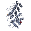
| Structure viewer | Molecule: MolmilJmol/JSmol |
|---|
- Downloads & links
-Download
| PDBx/mmCIF format | 4a8x.cif.gz | 150.8 KB | Display | PDBx/mmCIF format |
|---|---|---|---|---|
| PDB format | pdb4a8x.ent.gz | 120.2 KB | Display | PDB format |
| PDBx/mmJSON format | 4a8x.json.gz | Tree view | PDBx/mmJSON format | |
| Others |  Other downloads Other downloads |
-Validation report
| Arichive directory | https://data.pdbj.org/pub/pdb/validation_reports/a8/4a8xftp://data.pdbj.org/pub/pdb/validation_reports/a8/4a8x | HTTPS FTP |
|---|
-Related structure data
| Related structure data | 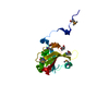 4a6qSC 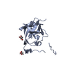 4a90C  1rk8S S: Starting model for refinement C: citing same article ( |
|---|---|
| Similar structure data |


-Links
 PDBj
PDBj


- Assembly
| Deposited unit | 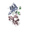
| ||||||||
|---|---|---|---|---|---|---|---|---|---|
| 1 |
| ||||||||
| Unit cell |
|
-Components
| #1: Protein | Mass: 10051.624 Da / Num. of mol.: 1 / Fragment: RRM DOMAIN, RESIDUES 122-207 Source method: isolated from a genetically manipulated source Source: (gene. exp.) HOMO SAPIENS (human) / Plasmid: PET SERIES / Production host:  |
|---|---|
| #2: Protein/peptide | Mass: 4707.469 Da / Num. of mol.: 1 / Fragment: RSB DOMAIN, RESIDUES 648-687 Source method: isolated from a genetically manipulated source Source: (gene. exp.) |
| #3: Protein | Mass: 14988.154 Da / Num. of mol.: 1 / Fragment: RESIDUES 14-143 Source method: isolated from a genetically manipulated source Source: (gene. exp.) |
| #4: Water | ChemComp-HOH /  Mass: 18.015 Da / Num. of mol.: 179 / Source method: isolated from a natural source / Formula: H2O Mass: 18.015 Da / Num. of mol.: 179 / Source method: isolated from a natural source / Formula: H2O |
| Sequence details | SM ARE RESIDUALS FROM TAG CLEAVAGE |
-Experimental details
-Experiment
| Experiment | Method: X-RAY DIFFRACTION / Number of used crystals: 1 |
|---|
- Sample preparation
| Crystal | Density Matthews: 2.28 Å3/Da / Density % sol: 46.13 % / Description: NONE |
|---|---|
| Crystal grow | pH: 7.5 / Details: 100 MM TRIS PH 7.5 25% PEG 3350 |
-Data collection
| Diffraction | Mean temperature: 100 K |
|---|---|
| Diffraction source | Source: SYNCHROTRON / Site: SLS  / Beamline: X06DA / Wavelength: 1 / Beamline: X06DA / Wavelength: 1 |
| Detector | Type: MARMOSAIC 225 mm CCD / Detector: CCD / Date: Jul 11, 2010 |
| Radiation | Protocol: SINGLE WAVELENGTH / Monochromatic (M) / Laue (L): M / Scattering type: x-ray |
| Radiation wavelength | Wavelength: 1 Å / Relative weight: 1 |
| Reflection | Resolution: 1.9→32.57 Å / Num. obs: 25760 / % possible obs: 98.9 % / Observed criterion σ(I): 3.8 / Redundancy: 7.5 % / Biso Wilson estimate: 32.27 Å2 / Rmerge(I) obs: 0.07 / Rsym value: 0.1 / Net I/σ(I): 14.4 |
| Reflection shell | Resolution: 1.9→2 Å / Redundancy: 7.5 % / Rmerge(I) obs: 0.4 / Mean I/σ(I) obs: 3.8 / Rsym value: 0.48 / % possible all: 100 |
- Processing
| Software |
| ||||||||||||||||||||||||||||||||||||||||||||||||||||||||||||||||||||||||||||||||||||||||||||||||||||
|---|---|---|---|---|---|---|---|---|---|---|---|---|---|---|---|---|---|---|---|---|---|---|---|---|---|---|---|---|---|---|---|---|---|---|---|---|---|---|---|---|---|---|---|---|---|---|---|---|---|---|---|---|---|---|---|---|---|---|---|---|---|---|---|---|---|---|---|---|---|---|---|---|---|---|---|---|---|---|---|---|---|---|---|---|---|---|---|---|---|---|---|---|---|---|---|---|---|---|---|---|---|
| Refinement | Method to determine structure: MOLECULAR REPLACEMENT Starting model: PDB ENTRIES 1RK8 AND 4A6Q Resolution: 1.9→36.289 Å / SU ML: 0.54 / σ(F): 0 / Phase error: 23.08 / Stereochemistry target values: ML Details: DISORDERED SIDE CHAIN WERE TRUNCATED AT C ALPHA. THE 96-100 LOOP OF SAP18 (CHAIN C) DISPLAYS POOR DENSITY AND WAS MODELED BASED ON ENTRY 4A6Q
| ||||||||||||||||||||||||||||||||||||||||||||||||||||||||||||||||||||||||||||||||||||||||||||||||||||
| Solvent computation | Shrinkage radii: 0.9 Å / VDW probe radii: 1.11 Å / Solvent model: FLAT BULK SOLVENT MODEL / Bsol: 41.834 Å2 / ksol: 0.309 e/Å3 | ||||||||||||||||||||||||||||||||||||||||||||||||||||||||||||||||||||||||||||||||||||||||||||||||||||
| Displacement parameters | Biso mean: 35 Å2
| ||||||||||||||||||||||||||||||||||||||||||||||||||||||||||||||||||||||||||||||||||||||||||||||||||||
| Refinement step | Cycle: LAST / Resolution: 1.9→36.289 Å
| ||||||||||||||||||||||||||||||||||||||||||||||||||||||||||||||||||||||||||||||||||||||||||||||||||||
| Refine LS restraints |
| ||||||||||||||||||||||||||||||||||||||||||||||||||||||||||||||||||||||||||||||||||||||||||||||||||||
| LS refinement shell |
| ||||||||||||||||||||||||||||||||||||||||||||||||||||||||||||||||||||||||||||||||||||||||||||||||||||
| Refinement TLS params. | Method: refined / Refine-ID: X-RAY DIFFRACTION
| ||||||||||||||||||||||||||||||||||||||||||||||||||||||||||||||||||||||||||||||||||||||||||||||||||||
| Refinement TLS group |
|
