 Movie
Movie Controller


+ Open data

- Basic information
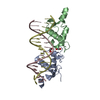
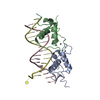
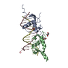
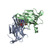
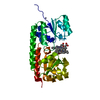
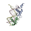
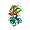
| Entry | Database: PDB / ID: 3g8x | ||||||
|---|---|---|---|---|---|---|---|
| Title | GR DNA binding domain:GilZ 16bp complex-65 | ||||||
 Components Components |
| ||||||
 Keywords Keywords | TRANSCRIPTION/DNA / glucocorticoid / DNA-binding / allostery / lever arm / transcription / hormone / Alternative initiation / Chromatin regulator / Cytoplasm / Lipid-binding / Metal-binding / Nucleus / Phosphoprotein / Polymorphism / Receptor / Steroid-binding / Transcription regulation / Ubl conjugation / Zinc / Zinc-finger / TRANSCRIPTION-DNA COMPLEX | ||||||
| Function / homology |  Function and homology information Function and homology informationnuclear receptor-mediated corticosteroid signaling pathway / negative regulation of behavioral fear response / muscle atrophy / HSP90 chaperone cycle for steroid hormone receptors (SHR) in the presence of ligand / negative regulation of synaptic plasticity / nuclear receptor-mediated glucocorticoid signaling pathway / positive regulation of nuclear receptor-mediated glucocorticoid signaling pathway / negative regulation of nuclear receptor-mediated glucocorticoid signaling pathway / response to inactivity / negative regulation of long-term synaptic depression ...nuclear receptor-mediated corticosteroid signaling pathway / negative regulation of behavioral fear response / muscle atrophy / HSP90 chaperone cycle for steroid hormone receptors (SHR) in the presence of ligand / negative regulation of synaptic plasticity / nuclear receptor-mediated glucocorticoid signaling pathway / positive regulation of nuclear receptor-mediated glucocorticoid signaling pathway / negative regulation of nuclear receptor-mediated glucocorticoid signaling pathway / response to inactivity / negative regulation of long-term synaptic depression / regulation of glucocorticoid biosynthetic process / nuclear glucocorticoid receptor activity / SUMOylation of intracellular receptors / positive regulation of cell growth involved in cardiac muscle cell development / Nuclear Receptor transcription pathway / response to mercury ion / steroid hormone binding / glucocorticoid metabolic process / response to cortisol / neuroinflammatory response / Leydig cell differentiation / mammary gland duct morphogenesis / microglia differentiation / cellular response to magnesium ion / astrocyte differentiation / response to arsenic-containing substance / maternal behavior / negative regulation of vascular permeability / regulation of gluconeogenesis / adrenal gland development / cellular response to glucocorticoid stimulus / cellular response to steroid hormone stimulus / response to dexamethasone / response to corticosterone / positive regulation of glutamate secretion / positive regulation of dendritic spine development / motor behavior / hormone binding / regulation of glucose metabolic process / nuclear receptor-mediated steroid hormone signaling pathway / estrogen response element binding / associative learning / response to electrical stimulus / cellular response to dexamethasone stimulus / cellular response to transforming growth factor beta stimulus / core promoter sequence-specific DNA binding / postsynaptic density, intracellular component / steroid binding / heat shock protein binding / lung development / Hsp70 protein binding / TBP-class protein binding / transcription initiation-coupled chromatin remodeling / positive regulation of cytokine production / response to activity / RNA polymerase II transcription regulatory region sequence-specific DNA binding / promoter-specific chromatin binding / female pregnancy / synaptic transmission, glutamatergic / circadian rhythm / Hsp90 protein binding / response to insulin / receptor tyrosine kinase binding / response to wounding / response to calcium ion / positive regulation of miRNA transcription / DNA-binding transcription repressor activity, RNA polymerase II-specific / nuclear receptor activity / spindle / sequence-specific double-stranded DNA binding / positive regulation of neuron apoptotic process / regulation of cell population proliferation / protein-containing complex assembly / double-stranded DNA binding / DNA-binding transcription activator activity, RNA polymerase II-specific / sequence-specific DNA binding / gene expression / dendritic spine / transcription coactivator activity / nuclear speck / RNA polymerase II cis-regulatory region sequence-specific DNA binding / chromatin remodeling / DNA-binding transcription factor activity / negative regulation of DNA-templated transcription / chromatin binding / regulation of transcription by RNA polymerase II / regulation of DNA-templated transcription / centrosome / protein kinase binding / negative regulation of apoptotic process / chromatin / protein-containing complex binding / glutamatergic synapse / negative regulation of transcription by RNA polymerase II / positive regulation of transcription by RNA polymerase II / protein-containing complex / mitochondrion / DNA binding / zinc ion binding / nucleoplasm Similarity search - Function | ||||||
| Biological species |  | ||||||
| Method |  X-RAY DIFFRACTION / SYNCHROTRON / MOLECULAR REPLACEMENT / Resolution: 2.05 Å X-RAY DIFFRACTION / SYNCHROTRON / MOLECULAR REPLACEMENT / Resolution: 2.05 Å | ||||||
 Authors Authors | Pufall, M.A. / Yamamoto, K.R. / Meijsing, S.H. | ||||||
 Citation Citation | Journal: Science / Year: 2009 Title: DNA binding site sequence directs glucocorticoid receptor structure and activity. Authors: Meijsing, S.H. / Pufall, M.A. / So, A.Y. / Bates, D.L. / Chen, L. / Yamamoto, K.R. | ||||||
| History |
|
- Structure visualization
| Structure viewer | Molecule: MolmilJmol/JSmol |
|---|
- Downloads & links
-Download
| PDBx/mmCIF format | 3g8x.cif.gz | 113.3 KB | Display | PDBx/mmCIF format |
|---|---|---|---|---|
| PDB format | pdb3g8x.ent.gz | 85.4 KB | Display | PDB format |
| PDBx/mmJSON format | 3g8x.json.gz | Tree view | PDBx/mmJSON format | |
| Others |  Other downloads Other downloads |
-Validation report
| Arichive directory | https://data.pdbj.org/pub/pdb/validation_reports/g8/3g8xftp://data.pdbj.org/pub/pdb/validation_reports/g8/3g8x | HTTPS FTP |
|---|
-Related structure data
| Related structure data |  3fylC  3g6pC  3g6qC  3g6rC  3g6tC  3g6uC  3g8uC  3g97C  3g99C  3g9iC  3g9jC  3g9mC 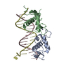 3g9oC  3g9pC  1gluS C: citing same article ( S: Starting model for refinement |
|---|---|
| Similar structure data |


-Links
 PDBj
PDBj


- Assembly
| Deposited unit | 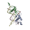
| ||||||||
|---|---|---|---|---|---|---|---|---|---|
| 1 |
| ||||||||
| Unit cell |
|
-Components
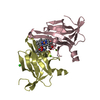
-Protein , 1 types, 2 molecules AB
| #1: Protein | Mass: 9962.758 Da / Num. of mol.: 2 / Fragment: UNP residues 440-525 Source method: isolated from a genetically manipulated source Source: (gene. exp.)  |
|---|
-DNA chain , 2 types, 2 molecules DC
| #2: DNA chain | Mass: 4873.178 Da / Num. of mol.: 1 / Source method: obtained synthetically / Details: oligonucleotide from Integrated DNA technologies |
|---|---|
| #3: DNA chain | Mass: 4922.216 Da / Num. of mol.: 1 / Source method: obtained synthetically / Details: oligonucleotide from Integrated DNA technologies |
-Non-polymers , 3 types, 73 molecules 


| #4: Chemical | ChemComp-ZN /  Mass: 65.409 Da / Num. of mol.: 4 / Source method: obtained synthetically / Formula: Zn Mass: 65.409 Da / Num. of mol.: 4 / Source method: obtained synthetically / Formula: Zn#5: Chemical |  Mass: 62.068 Da / Num. of mol.: 2 / Source method: obtained synthetically / Formula: C2H6O2 Mass: 62.068 Da / Num. of mol.: 2 / Source method: obtained synthetically / Formula: C2H6O2#6: Water | ChemComp-HOH / | Mass: 18.015 Da / Num. of mol.: 67 / Source method: isolated from a natural source / Formula: H2O |
|---|
-Details
| Has protein modification | Y |
|---|
-Experimental details
-Experiment
| Experiment | Method: X-RAY DIFFRACTION / Number of used crystals: 1 |
|---|
- Sample preparation
| Crystal | Density Matthews: 3.03 Å3/Da / Density % sol: 59.45 % | ||||||||||||||||||||||||||||||||
|---|---|---|---|---|---|---|---|---|---|---|---|---|---|---|---|---|---|---|---|---|---|---|---|---|---|---|---|---|---|---|---|---|---|
| Crystal grow | Temperature: 298 K / Method: vapor diffusion, hanging drop / pH: 7 Details: 50mM Na Cacodylate, pH 7.0, 2.25mM Spermine, 9mM MgCl2, 1.8 CoCl2, 5% PEG 400, VAPOR DIFFUSION, HANGING DROP, temperature 298K | ||||||||||||||||||||||||||||||||
| Components of the solutions |
|
-Data collection
| Diffraction | Mean temperature: 100 K |
|---|---|
| Diffraction source | Source: SYNCHROTRON / Site: ALS  / Beamline: 8.3.1 / Wavelength: 1.115869 Å / Beamline: 8.3.1 / Wavelength: 1.115869 Å |
| Detector | Type: ADSC QUANTUM 210 / Detector: CCD / Date: Apr 26, 2007 |
| Radiation | Monochromator: Double flat crystal, Si(111) / Protocol: SINGLE WAVELENGTH / Monochromatic (M) / Laue (L): M / Scattering type: x-ray |
| Radiation wavelength | Wavelength: 1.115869 Å / Relative weight: 1 |
| Reflection | Resolution: 2.05→40 Å / Num. obs: 21522 / % possible obs: 94.5 % / Observed criterion σ(I): 0 / Redundancy: 6.4 % / Biso Wilson estimate: 41.139 Å2 / Rmerge(I) obs: 0.137 / Rsym value: 0.183 / Net I/σ(I): 15 |
| Reflection shell | Resolution: 2.05→2.12 Å / Redundancy: 3.2 % / Rmerge(I) obs: 0.456 / Mean I/σ(I) obs: 2.2 / Num. unique all: 1604 / Rsym value: 0.659 / % possible all: 71.1 |
- Processing
| Software |
| |||||||||||||||||||||||||||||||||||||||||||||||||||||||||||||||||||||||||||||||||||||||||||||||||||||||||||||||||||||||||||||
|---|---|---|---|---|---|---|---|---|---|---|---|---|---|---|---|---|---|---|---|---|---|---|---|---|---|---|---|---|---|---|---|---|---|---|---|---|---|---|---|---|---|---|---|---|---|---|---|---|---|---|---|---|---|---|---|---|---|---|---|---|---|---|---|---|---|---|---|---|---|---|---|---|---|---|---|---|---|---|---|---|---|---|---|---|---|---|---|---|---|---|---|---|---|---|---|---|---|---|---|---|---|---|---|---|---|---|---|---|---|---|---|---|---|---|---|---|---|---|---|---|---|---|---|---|---|---|
| Refinement | Method to determine structure: MOLECULAR REPLACEMENT Starting model: PDB entry 1GLU Resolution: 2.05→35.363 Å / SU ML: 2.04 / σ(F): 0.38 / Stereochemistry target values: ML
| |||||||||||||||||||||||||||||||||||||||||||||||||||||||||||||||||||||||||||||||||||||||||||||||||||||||||||||||||||||||||||||
| Solvent computation | Shrinkage radii: 0.9 Å / VDW probe radii: 1.11 Å / Solvent model: FLAT BULK SOLVENT MODEL / Bsol: 60.89 Å2 / ksol: 0.311 e/Å3 | |||||||||||||||||||||||||||||||||||||||||||||||||||||||||||||||||||||||||||||||||||||||||||||||||||||||||||||||||||||||||||||
| Displacement parameters | Biso mean: 84.4 Å2 | |||||||||||||||||||||||||||||||||||||||||||||||||||||||||||||||||||||||||||||||||||||||||||||||||||||||||||||||||||||||||||||
| Refinement step | Cycle: LAST / Resolution: 2.05→35.363 Å
| |||||||||||||||||||||||||||||||||||||||||||||||||||||||||||||||||||||||||||||||||||||||||||||||||||||||||||||||||||||||||||||
| Refine LS restraints |
| |||||||||||||||||||||||||||||||||||||||||||||||||||||||||||||||||||||||||||||||||||||||||||||||||||||||||||||||||||||||||||||
| LS refinement shell | Refine-ID: X-RAY DIFFRACTION
| |||||||||||||||||||||||||||||||||||||||||||||||||||||||||||||||||||||||||||||||||||||||||||||||||||||||||||||||||||||||||||||
| Refinement TLS params. | Method: refined / Refine-ID: X-RAY DIFFRACTION
| |||||||||||||||||||||||||||||||||||||||||||||||||||||||||||||||||||||||||||||||||||||||||||||||||||||||||||||||||||||||||||||
| Refinement TLS group |
|
