[English] 日本語
Yorodumi
- PDB-4a2s: Structure of the engineered retro-aldolase RA95.5 -

+
Open data


ID or keywords:

Loading...

-
Basic information

Entry
Database: PDB / ID: 4a2s
TitleStructure of the engineered retro-aldolase RA95.5
ComponentsINDOLE-3-GLYCEROL PHOSPHATE SYNTHASE
KeywordsLYASE / ENGINEERED ENZYME / DIRECTED EVOLUTION
Function / homology
Function and homology information


indole-3-glycerol-phosphate synthase / indole-3-glycerol-phosphate synthase activity / phosphoribosylanthranilate isomerase activity / tryptophan biosynthetic process
Similarity search - Function
Indole-3-glycerol phosphate synthase / Indole-3-glycerol phosphate synthase, conserved site / Indole-3-glycerol phosphate synthase signature. / Indole-3-glycerol phosphate synthase domain / Indole-3-glycerol phosphate synthase / Ribulose-phosphate binding barrel / Aldolase class I / Aldolase-type TIM barrel / TIM Barrel / Alpha-Beta Barrel / Alpha Beta
Similarity search - Domain/homology
1-(6-METHOXYNAPHTHALEN-2-YL)BUTANE-1,3-DIONE / Indole-3-glycerol phosphate synthase
Similarity search - Component
Biological speciesSULFOLOBUS SOLFATARICUS (archaea)
MethodX-RAY DIFFRACTION / SYNCHROTRON / MOLECULAR REPLACEMENT / Resolution: 1.4 Å
AuthorsGiger, L. / Caner, S. / Kast, P. / Baker, D. / Ban, N. / Hilvert, D.
CitationJournal: Nat.Chem.Biol. / Year: 2013
Title: Evolution of a designed retro-aldolase leads to complete active site remodeling.
Authors: Giger, L. / Caner, S. / Obexer, R. / Kast, P. / Baker, D. / Ban, N. / Hilvert, D.
History
DepositionSep 28, 2011Deposition site: PDBE / Processing site: PDBE
Revision 1.0Nov 7, 2012Provider: repository / Type: Initial release
Revision 1.1Nov 21, 2012Group: Atomic model
Revision 1.2Feb 6, 2013Group: Atomic model / Other
Revision 1.3Jun 12, 2013Group: Database references
Revision 1.4Jun 19, 2013Group: Database references
Revision 1.5Jul 31, 2013Group: Database references
Revision 2.0May 15, 2019Group: Atomic model / Data collection ...Atomic model / Data collection / Database references / Derived calculations / Experimental preparation / Other
Category: atom_site / citation ...atom_site / citation / database_PDB_rev / database_PDB_rev_record / exptl_crystal_grow / pdbx_database_proc / pdbx_database_status / struct_biol / struct_conn
Item: _atom_site.B_iso_or_equiv / _atom_site.Cartn_x ..._atom_site.B_iso_or_equiv / _atom_site.Cartn_x / _atom_site.Cartn_y / _atom_site.Cartn_z / _atom_site.pdbx_auth_atom_name / _citation.journal_id_ISSN / _citation.page_last / _citation.pdbx_database_id_DOI / _citation.title / _exptl_crystal_grow.method / _exptl_crystal_grow.temp / _pdbx_database_status.recvd_author_approval / _struct_conn.pdbx_leaving_atom_flag
Revision 2.1Dec 20, 2023Group: Data collection / Database references ...Data collection / Database references / Derived calculations / Other / Refinement description
Category: chem_comp_atom / chem_comp_bond ...chem_comp_atom / chem_comp_bond / database_2 / pdbx_database_status / pdbx_initial_refinement_model / struct_site
Item: _database_2.pdbx_DOI / _database_2.pdbx_database_accession ..._database_2.pdbx_DOI / _database_2.pdbx_database_accession / _pdbx_database_status.status_code_sf / _struct_site.pdbx_auth_asym_id / _struct_site.pdbx_auth_comp_id / _struct_site.pdbx_auth_seq_id
Remark 700 
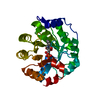
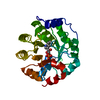
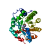
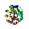
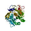
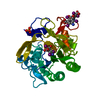
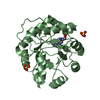
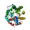
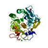
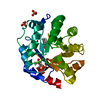
SHEET DETERMINATION METHOD: DSSP THE SHEETS PRESENTED AS "AA" IN EACH CHAIN ON SHEET RECORDS BELOW ... SHEET DETERMINATION METHOD: DSSP THE SHEETS PRESENTED AS "AA" IN EACH CHAIN ON SHEET RECORDS BELOW IS ACTUALLY AN 8-STRANDED BARREL THIS IS REPRESENTED BY A 9-STRANDED SHEET IN WHICH THE FIRST AND LAST STRANDS ARE IDENTICAL.

-
Structure visualization

Structure viewerMolecule:
MolmilJmol/JSmol

Downloads & links

-
Assembly

Deposited unit
A: INDOLE-3-GLYCEROL PHOSPHATE SYNTHASE
hetero molecules


Theoretical massNumber of molelcules
Total (without water)30,2343
Polymers29,7491
Non-polymers4852
Water3,747208
1


  • Idetical with deposited unit
  • defined by author&software
TypeNameSymmetry operationNumber
identity operation1_555x,y,z1
MethodPISA
Unit cell
Length a, b, c (Å)53.810, 62.810, 93.210
Angle α, β, γ (deg.)90.00, 90.00, 90.00
Int Tables number19
Space group name H-MP212121

-
Components

#1: Protein INDOLE-3-GLYCEROL PHOSPHATE SYNTHASE / IGPS / ENGINEERED RETRO-ALDOL ENZYME RA95.5-5


Mass: 29749.297 Da / Num. of mol.: 1 / Fragment: TIM-BARREL FOLD, RESIDUES 1-245 / Mutation: YES
Source method: isolated from a genetically manipulated source
Details: THE SEQUENCE WAS DESIGNED BASED ON INDOLE-3-GLYCEROL PHOSPHATE SYNTHASE NATURALLY FOUND IN SULFOLOBUS SOLFATARICUS.
Source: (gene. exp.) SULFOLOBUS SOLFATARICUS (archaea) / Production host: ESCHERICHIA COLI (E. coli) / Strain (production host): BL21(DE3) / Variant (production host): GOLD
References: UniProt: Q06121, indole-3-glycerol-phosphate synthase
#2: Chemical ChemComp-3NK / 1-(6-METHOXYNAPHTHALEN-2-YL)BUTANE-1,3-DIONE


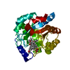
Mass: 242.270 Da / Num. of mol.: 2 / Source method: obtained synthetically / Formula: C15H14O3
#3: Water ChemComp-HOH / water / Water


Mass: 18.015 Da / Num. of mol.: 208 / Source method: isolated from a natural source / Formula: H2O
Compound detailsENGINEERED RESIDUE IN CHAIN A, LYS 10 TO GLU ENGINEERED RESIDUE IN CHAIN A, PHE 22 TO VAL ...ENGINEERED RESIDUE IN CHAIN A, LYS 10 TO GLU ENGINEERED RESIDUE IN CHAIN A, PHE 22 TO VAL ENGINEERED RESIDUE IN CHAIN A, GLU 51 TO TYR ENGINEERED RESIDUE IN CHAIN A, LYS 53 TO SER ENGINEERED RESIDUE IN CHAIN A, LEU 83 TO LYS ENGINEERED RESIDUE IN CHAIN A, LYS 110 TO SER ENGINEERED RESIDUE IN CHAIN A, GLU 159 TO LEU ENGINEERED RESIDUE IN CHAIN A, ASN 180 TO PHE ENGINEERED RESIDUE IN CHAIN A, ARG 182 TO MET ENGINEERED RESIDUE IN CHAIN A, ASP 183 TO ASN ENGINEERED RESIDUE IN CHAIN A, LEU 184 TO PHE ENGINEERED RESIDUE IN CHAIN A, GLU 210 TO LYS ENGINEERED RESIDUE IN CHAIN A, SER 211 TO LEU ENGINEERED RESIDUE IN CHAIN A, GLY 233 TO SER

-
Experimental details

-
Experiment

ExperimentMethod: X-RAY DIFFRACTION / Number of used crystals: 1

-
Sample preparation

CrystalDensity Matthews: 2.7 Å3/Da / Density % sol: 54.2 % / Description: NONE
Crystal growTemperature: 301 K / Method: vapor diffusion
Details: 0.1 M HEPES KOH PH 7.6, 23% W/V PEG 3350,20 MM NA2HPO4, 28 DEG. CELSIUS.

-
Data collection

DiffractionMean temperature: 100 K
Diffraction sourceSource: SYNCHROTRON / Site: SLS / Beamline: X06SA / Wavelength: 1
DetectorType: DECTRIS PILATUS 6M / Detector: PIXEL / Date: Sep 14, 2010
RadiationProtocol: SINGLE WAVELENGTH / Monochromatic (M) / Laue (L): M / Scattering type: x-ray
Radiation wavelengthWavelength: 1 Å / Relative weight: 1
ReflectionResolution: 1.4→150 Å / Num. obs: 62630 / % possible obs: 99.3 % / Observed criterion σ(I): -3 / Redundancy: 6.5 % / Biso Wilson estimate: 22.79 Å2 / Rmerge(I) obs: 0.02 / Net I/σ(I): 31.12
Reflection shellResolution: 1.4→1.44 Å / Redundancy: 2.6 % / Rmerge(I) obs: 0.87 / Mean I/σ(I) obs: 2.45 / % possible all: 95.1

-
Processing

Software
NameVersionClassification
PHENIX(PHENIX.REFINE)refinement
XDSdata reduction
XSCALEdata scaling
PHASERphasing
RefinementMethod to determine structure: MOLECULAR REPLACEMENT
Starting model: PDB ENTRY 1LBL
Resolution: 1.4→7.998 Å / SU ML: 0.25 / σ(F): 2 / Phase error: 14.55 / Stereochemistry target values: ML
RfactorNum. reflection% reflection
Rfree0.1616 1866 3 %
Rwork0.1319 --
obs0.1328 62213 99.52 %
Solvent computationShrinkage radii: 0.05 Å / VDW probe radii: 0.3 Å / Solvent model: FLAT BULK SOLVENT MODEL / Bsol: 92.553 Å2 / ksol: 0.6 e/Å3
Displacement parametersBiso mean: 38.2 Å2
Baniso -1Baniso -2Baniso -3
1-2.0873 Å20 Å20 Å2
2---3.2544 Å20 Å2
3---0.5936 Å2
Refinement stepCycle: LAST / Resolution: 1.4→7.998 Å
ProteinNucleic acidLigandSolventTotal
Num. atoms1995 0 34 208 2237
Refine LS restraints
Refine-IDTypeDev idealNumber
X-RAY DIFFRACTIONf_bond_d0.0132296
X-RAY DIFFRACTIONf_angle_d1.5463125
X-RAY DIFFRACTIONf_dihedral_angle_d17.17923
X-RAY DIFFRACTIONf_chiral_restr0.113350
X-RAY DIFFRACTIONf_plane_restr0.007408
LS refinement shell
Resolution (Å)Rfactor RfreeNum. reflection RfreeRfactor RworkNum. reflection RworkRefine-ID% reflection obs (%)
1.4-1.43760.2811390.23864499X-RAY DIFFRACTION98
1.4376-1.47970.25781420.20324573X-RAY DIFFRACTION100
1.4797-1.52710.23251410.17554599X-RAY DIFFRACTION100
1.5271-1.58130.16391430.14574633X-RAY DIFFRACTION100
1.5813-1.64410.15981420.1284581X-RAY DIFFRACTION100
1.6441-1.71820.15931430.11824618X-RAY DIFFRACTION100
1.7182-1.80780.15321430.10534631X-RAY DIFFRACTION100
1.8078-1.91960.14491450.10014674X-RAY DIFFRACTION100
1.9196-2.06550.14431420.09984603X-RAY DIFFRACTION99
2.0655-2.2690.12581440.10534669X-RAY DIFFRACTION100
2.269-2.58760.14141460.11134700X-RAY DIFFRACTION100
2.5876-3.22410.17481450.13224690X-RAY DIFFRACTION99
3.2241-7.99830.16631510.1514877X-RAY DIFFRACTION100

+
About Yorodumi

-
News

-
Feb 9, 2022. New format data for meta-information of EMDB entries

New format data for meta-information of EMDB entries

  • Version 3 of the EMDB header file is now the official format.
  • The previous official version 1.9 will be removed from the archive.

Related info.:EMDB header

External links:wwPDB to switch to version 3 of the EMDB data model

-
Aug 12, 2020. Covid-19 info

Covid-19 info

URL: https://pdbj.org/emnavi/covid19.php

New page: Covid-19 featured information page in EM Navigator.

Related info.:Covid-19 info / Mar 5, 2020. Novel coronavirus structure data

+
Mar 5, 2020. Novel coronavirus structure data

Novel coronavirus structure data

Related info.:Yorodumi Speices / Aug 12, 2020. Covid-19 info

External links:COVID-19 featured content - PDBj / Molecule of the Month (242):Coronavirus Proteases

+
Jan 31, 2019. EMDB accession codes are about to change! (news from PDBe EMDB page)

EMDB accession codes are about to change! (news from PDBe EMDB page)

  • The allocation of 4 digits for EMDB accession codes will soon come to an end. Whilst these codes will remain in use, new EMDB accession codes will include an additional digit and will expand incrementally as the available range of codes is exhausted. The current 4-digit format prefixed with “EMD-” (i.e. EMD-XXXX) will advance to a 5-digit format (i.e. EMD-XXXXX), and so on. It is currently estimated that the 4-digit codes will be depleted around Spring 2019, at which point the 5-digit format will come into force.
  • The EM Navigator/Yorodumi systems omit the EMD- prefix.

Related info.:Q: What is EMD? / ID/Accession-code notation in Yorodumi/EM Navigator

External links:EMDB Accession Codes are Changing Soon! / Contact to PDBj

+
Jul 12, 2017. Major update of PDB

Major update of PDB

  • wwPDB released updated PDB data conforming to the new PDBx/mmCIF dictionary.
  • This is a major update changing the version number from 4 to 5, and with Remediation, in which all the entries are updated.
  • In this update, many items about electron microscopy experimental information are reorganized (e.g. em_software).
  • Now, EM Navigator and Yorodumi are based on the updated data.

External links:wwPDB Remediation / Enriched Model Files Conforming to OneDep Data Standards Now Available in the PDB FTP Archive

-
Yorodumi

Thousand views of thousand structures

  • Yorodumi is a browser for structure data from EMDB, PDB, SASBDB, etc.
  • This page is also the successor to EM Navigator detail page, and also detail information page/front-end page for Omokage search.
  • The word "yorodu" (or yorozu) is an old Japanese word meaning "ten thousand". "mi" (miru) is to see.

Related info.:EMDB / PDB / SASBDB / Comparison of 3 databanks / Yorodumi Search / Aug 31, 2016. New EM Navigator & Yorodumi / Yorodumi Papers / Jmol/JSmol / Function and homology information / Changes in new EM Navigator and Yorodumi

Read more