 Movie
Movie Controller

[English] 日本語
 Yorodumi
- PDB-1lbl: Crystal structure of indole-3-glycerol phosphate synthase (IGPS) ... -
+ Open data

- Basic information
| Entry | Database: PDB / ID: 1lbl | ||||||
|---|---|---|---|---|---|---|---|
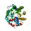
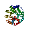
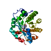
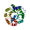
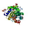
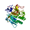
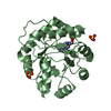
| Title | Crystal structure of indole-3-glycerol phosphate synthase (IGPS) in complex with 1-(o-carboxyphenylamino)-1-deoxyribulose 5'-phosphate (CdRP) | ||||||
 Components Components | indole-3-glycerol phosphate synthase | ||||||
 Keywords Keywords | LYASE / BETA BARREL / tryptophan biosynthesis / protein ligand complex / substrate complex | ||||||
| Function / homology |  Function and homology information Function and homology informationindole-3-glycerol-phosphate synthase / indole-3-glycerol-phosphate synthase activity / L-tryptophan biosynthetic process Similarity search - Function | ||||||
| Biological species |   Sulfolobus solfataricus (archaea) Sulfolobus solfataricus (archaea) | ||||||
| Method |  X-RAY DIFFRACTION / FOURIER SYNTHESIS / Resolution: 2.4 Å X-RAY DIFFRACTION / FOURIER SYNTHESIS / Resolution: 2.4 Å | ||||||
 Authors Authors | Hennig, M. / Darimont, B.D. / Kirschner, K. / Jansonius, J.N. | ||||||
 Citation Citation | Journal: J.Mol.Biol. / Year: 2002 Title: The catalytic mechanism of indole-3-glycerol phosphate synthase: Crystal structures of complexes of the enzyme from Sulfolobus solfataricus with the substrate analogue, substrate, and product Authors: Hennig, M. / Darimont, B.D. / Jansonius, J.N. / Kirschner, K. #1: Journal: Structure / Year: 1995Title: 2.0 A structure of indole-3-glycerol phosphate synthase from the hyperthermophile Sulfolobus solfataricus: possible determinants of protein stability. | ||||||
| History |
|
- Structure visualization
| Structure viewer | Molecule: MolmilJmol/JSmol |
|---|
- Downloads & links
-Download
| PDBx/mmCIF format | 1lbl.cif.gz | 67.8 KB | Display | PDBx/mmCIF format |
|---|---|---|---|---|
| PDB format | pdb1lbl.ent.gz | 49.1 KB | Display | PDB format |
| PDBx/mmJSON format | 1lbl.json.gz | Tree view | PDBx/mmJSON format | |
| Others |  Other downloads Other downloads |
-Validation report
| Arichive directory | https://data.pdbj.org/pub/pdb/validation_reports/lb/1lblftp://data.pdbj.org/pub/pdb/validation_reports/lb/1lbl | HTTPS FTP |
|---|
-Related structure data
| Related structure data | 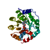 1a53C 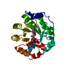 1lbfC  1igsS S: Starting model for refinement C: citing same article ( |
|---|---|
| Similar structure data |


-Links
 PDBj
PDBj
- Assembly
| Deposited unit | 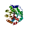
| ||||||||
|---|---|---|---|---|---|---|---|---|---|
| 1 |
| ||||||||
| Unit cell |
|
-Components
| #1: Protein | Mass: 28494.906 Da / Num. of mol.: 1 Source method: isolated from a genetically manipulated source Source: (gene. exp.) Sulfolobus solfataricus (archaea) / Production host:  References: UniProt: Q06121, indole-3-glycerol-phosphate synthase |
|---|---|
| #2: Chemical | ChemComp-137 /   Mass: 351.246 Da / Num. of mol.: 1 / Source method: obtained synthetically / Formula: C12H18NO9P Mass: 351.246 Da / Num. of mol.: 1 / Source method: obtained synthetically / Formula: C12H18NO9P |
| #3: Water | ChemComp-HOH /  Mass: 18.015 Da / Num. of mol.: 203 / Source method: isolated from a natural source / Formula: H2O Mass: 18.015 Da / Num. of mol.: 203 / Source method: isolated from a natural source / Formula: H2O |
-Experimental details
-Experiment
| Experiment | Method: X-RAY DIFFRACTION / Number of used crystals: 1 |
|---|
- Sample preparation
| Crystal | Density Matthews: 3.91 Å3/Da / Density % sol: 68.5 % |
|---|---|
| Crystal grow | Temperature: 279 K / Method: vapor diffusion, hanging drop / pH: 6.5 Details: 1.2M ammonium sulphate, 50 mM potassium phosphate buffer, pH 6.5, VAPOR DIFFUSION, HANGING DROP, temperature 279K |
-Data collection
| Diffraction | Mean temperature: 273 K |
|---|---|
| Diffraction source | Source: ROTATING ANODE / Type: ELLIOTT GX-20 / Wavelength: 1.5418 Å |
| Detector | Type: MARRESEARCH / Detector: IMAGE PLATE / Date: Sep 18, 1995 |
| Radiation | Monochromator: graphite / Protocol: SINGLE WAVELENGTH / Monochromatic (M) / Laue (L): M / Scattering type: x-ray |
| Radiation wavelength | Wavelength: 1.5418 Å / Relative weight: 1 |
| Reflection | Resolution: 2.4→15 Å / Num. all: 60396 / Num. obs: 17120 / % possible obs: 94.6 % / Observed criterion σ(F): 0 / Observed criterion σ(I): 0 / Rmerge(I) obs: 0.106 |
- Processing
| Software |
| ||||||||||||||||||||
|---|---|---|---|---|---|---|---|---|---|---|---|---|---|---|---|---|---|---|---|---|---|
| Refinement | Method to determine structure: FOURIER SYNTHESIS Starting model: 1IGS Resolution: 2.4→15 Å / Cross valid method: THROUGHOUT / σ(F): 0 / Stereochemistry target values: Engh & Huber
| ||||||||||||||||||||
| Refinement step | Cycle: LAST / Resolution: 2.4→15 Å
| ||||||||||||||||||||
| Refine LS restraints |
|
