 Movie
Movie Controller

[English] 日本語
 Yorodumi
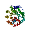
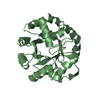
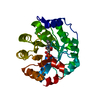
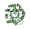
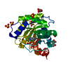
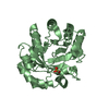
- PDB-1a53: COMPLEX OF INDOLE-3-GLYCEROLPHOSPHATE SYNTHASE FROM SULFOLOBUS SO... -
+ Open data

- Basic information
| Entry | Database: PDB / ID: 1a53 | ||||||
|---|---|---|---|---|---|---|---|
| Title | COMPLEX OF INDOLE-3-GLYCEROLPHOSPHATE SYNTHASE FROM SULFOLOBUS SOLFATARICUS WITH INDOLE-3-GLYCEROLPHOSPHATE AT 2.0 A RESOLUTION | ||||||
 Components Components | INDOLE-3-GLYCEROLPHOSPHATE SYNTHASE | ||||||
 Keywords Keywords | SYNTHASE / THERMOSTABLE / TIM-BARREL | ||||||
| Function / homology |  Function and homology information Function and homology informationindole-3-glycerol-phosphate synthase / indole-3-glycerol-phosphate synthase activity / L-tryptophan biosynthetic process Similarity search - Function | ||||||
| Biological species |   Sulfolobus solfataricus (archaea) Sulfolobus solfataricus (archaea) | ||||||
| Method |  X-RAY DIFFRACTION / MOLECULAR REPLACEMENT / Resolution: 2 Å X-RAY DIFFRACTION / MOLECULAR REPLACEMENT / Resolution: 2 Å | ||||||
 Authors Authors | Hennig, M. / Darimont, B. / Kirschner, K. / Jansonius, J.N. | ||||||
 Citation Citation | Journal: J.Mol.Biol. / Year: 2002 Title: The catalytic mechanism of indole-3-glycerol phosphate synthase: crystal structures of complexes of the enzyme from Sulfolobus solfataricus with substrate analogue, substrate, and product. Authors: Hennig, M. / Darimont, B.D. / Jansonius, J.N. / Kirschner, K. #1: Journal: Structure / Year: 1995Title: 2.0 A Structure of Indole-3-Glycerol Phosphate Synthase from the Hyperthermophile Sulfolobus Solfataricus: Possible Determinants of Protein Stability Authors: Hennig, M. / Darimont, B. / Sterner, R. / Kirschner, K. / Jansonius, J.N. | ||||||
| History |
|
- Structure visualization
| Structure viewer | Molecule: MolmilJmol/JSmol |
|---|
- Downloads & links
-Download
| PDBx/mmCIF format | 1a53.cif.gz | 68.4 KB | Display | PDBx/mmCIF format |
|---|---|---|---|---|
| PDB format | pdb1a53.ent.gz | 50.1 KB | Display | PDB format |
| PDBx/mmJSON format | 1a53.json.gz | Tree view | PDBx/mmJSON format | |
| Others |  Other downloads Other downloads |
-Validation report
| Arichive directory | https://data.pdbj.org/pub/pdb/validation_reports/a5/1a53ftp://data.pdbj.org/pub/pdb/validation_reports/a5/1a53 | HTTPS FTP |
|---|
-Related structure data
| Related structure data | 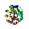 1lbfC  1lblC  1igsS S: Starting model for refinement C: citing same article ( |
|---|---|
| Similar structure data |


-Links
 PDBj
PDBj
- Assembly
| Deposited unit | 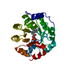
| ||||||||
|---|---|---|---|---|---|---|---|---|---|
| 1 |
| ||||||||
| Unit cell |
|
-Components
| #1: Protein | Mass: 28494.906 Da / Num. of mol.: 1 Source method: isolated from a genetically manipulated source Source: (gene. exp.) Sulfolobus solfataricus (archaea) / Gene: TRPC / Plasmid: REPRESSOR PLASMID PDM / Species (production host): Escherichia coli / Gene (production host): TRPCProduction host:  Strain (production host): W3110 References: UniProt: Q06121, indole-3-glycerol-phosphate synthase |
|---|---|
| #2: Chemical | ChemComp-IGP /   Mass: 287.206 Da / Num. of mol.: 1 / Source method: obtained synthetically / Formula: C11H14NO6P Mass: 287.206 Da / Num. of mol.: 1 / Source method: obtained synthetically / Formula: C11H14NO6P |
| #3: Water | ChemComp-HOH /  Mass: 18.015 Da / Num. of mol.: 242 / Source method: isolated from a natural source / Formula: H2O Mass: 18.015 Da / Num. of mol.: 242 / Source method: isolated from a natural source / Formula: H2O |
-Experimental details
-Experiment
| Experiment | Method: X-RAY DIFFRACTION / Number of used crystals: 1 |
|---|
- Sample preparation
| Crystal | Density Matthews: 3.91 Å3/Da / Density % sol: 68.5 % | ||||||||||||||||||||
|---|---|---|---|---|---|---|---|---|---|---|---|---|---|---|---|---|---|---|---|---|---|
| Crystal grow | pH: 5.5 / Details: pH 5.5 | ||||||||||||||||||||
| Crystal grow | *PLUS Temperature: 4 ℃ / Method: vapor diffusion, hanging drop | ||||||||||||||||||||
| Components of the solutions | *PLUS
|
-Data collection
| Diffraction | Mean temperature: 277 K |
|---|---|
| Diffraction source | Source: ROTATING ANODE / Type: ELLIOTT GX-21 / Wavelength: 1.5418 |
| Detector | Type: MARRESEARCH / Detector: IMAGE PLATE / Date: Aug 10, 1994 |
| Radiation | Monochromator: GRAPHITE(002) / Monochromatic (M) / Laue (L): M / Scattering type: x-ray |
| Radiation wavelength | Wavelength: 1.5418 Å / Relative weight: 1 |
| Reflection | Resolution: 2→15 Å / Num. obs: 27283 / % possible obs: 91.1 % / Observed criterion σ(I): 0 / Redundancy: 5.1 % / Biso Wilson estimate: 32.2 Å2 / Rmerge(I) obs: 0.063 / Net I/σ(I): 10 |
| Reflection shell | Resolution: 2→2.05 Å / Redundancy: 4.4 % / Rmerge(I) obs: 0.39 / Mean I/σ(I) obs: 1.6 / Rsym value: 0.39 / % possible all: 48 |
| Reflection | *PLUS Num. obs: 28014 / Num. measured all: 141730 |
- Processing
| Software |
| ||||||||||||||||||||||||||||||||||||||||||||||||||||||||||||
|---|---|---|---|---|---|---|---|---|---|---|---|---|---|---|---|---|---|---|---|---|---|---|---|---|---|---|---|---|---|---|---|---|---|---|---|---|---|---|---|---|---|---|---|---|---|---|---|---|---|---|---|---|---|---|---|---|---|---|---|---|---|
| Refinement | Method to determine structure: MOLECULAR REPLACEMENT Starting model: 1IGS Resolution: 2→15 Å / Data cutoff low absF: 0 / Isotropic thermal model: RESTRAINED / Cross valid method: THROUGHOUT / σ(F): 0
| ||||||||||||||||||||||||||||||||||||||||||||||||||||||||||||
| Displacement parameters | Biso mean: 29.6 Å2 | ||||||||||||||||||||||||||||||||||||||||||||||||||||||||||||
| Refinement step | Cycle: LAST / Resolution: 2→15 Å
| ||||||||||||||||||||||||||||||||||||||||||||||||||||||||||||
| Refine LS restraints |
| ||||||||||||||||||||||||||||||||||||||||||||||||||||||||||||
| LS refinement shell | Resolution: 2→2.07 Å / Total num. of bins used: 10
| ||||||||||||||||||||||||||||||||||||||||||||||||||||||||||||
| Xplor file |
|
