[English] 日本語
Yorodumi
- PDB-4a2p: Structure of duck RIG-I helicase domain -

+
Open data


ID or keywords:

Loading...

-
Basic information

Entry
Database: PDB / ID: 4a2p
TitleStructure of duck RIG-I helicase domain
ComponentsRETINOIC ACID INDUCIBLE PROTEIN I
KeywordsHYDROLASE / SUPERFAMILY 2 RNA HELICASE / ATP AND DSRNA BINDING / ANTIVIRAL SIGNALLING PATHWAY
Function / homology
Function and homology information


antiviral innate immune response / double-stranded RNA binding / RNA helicase activity / single-stranded RNA binding / hydrolase activity / RNA helicase / RNA binding / zinc ion binding / ATP binding / cytoplasm
Similarity search - Function
phosphoenolpyruvate carboxylase, domain 3 - #30 / phosphoenolpyruvate carboxylase, domain 3 / RIG-I, CARD domain repeat 2 / RIG-I-like receptor, C-terminal / RIG-I receptor C-terminal domain / RIG-I-like receptor, C-terminal regulatory domain / RIG-I-like receptor, C-terminal domain superfamily / C-terminal domain of RIG-I / RIG-I-like receptor (RLR) C-terminal regulatory (CTR) domain profile. / Caspase recruitment domain ...phosphoenolpyruvate carboxylase, domain 3 - #30 / phosphoenolpyruvate carboxylase, domain 3 / RIG-I, CARD domain repeat 2 / RIG-I-like receptor, C-terminal / RIG-I receptor C-terminal domain / RIG-I-like receptor, C-terminal regulatory domain / RIG-I-like receptor, C-terminal domain superfamily / C-terminal domain of RIG-I / RIG-I-like receptor (RLR) C-terminal regulatory (CTR) domain profile. / Caspase recruitment domain / Caspase recruitment domain / Death-like domain superfamily / DEAD/DEAH box helicase / DEAD/DEAH box helicase domain / Helicase conserved C-terminal domain / helicase superfamily c-terminal domain / Superfamilies 1 and 2 helicase C-terminal domain profile. / Superfamilies 1 and 2 helicase ATP-binding type-1 domain profile. / DEAD-like helicases superfamily / Helicase, C-terminal / Helicase superfamily 1/2, ATP-binding domain / P-loop containing nucleotide triphosphate hydrolases / Up-down Bundle / P-loop containing nucleoside triphosphate hydrolase / Rossmann fold / 3-Layer(aba) Sandwich / Mainly Alpha / Alpha Beta
Similarity search - Domain/homology
Biological speciesANAS PLATYRHYNCHOS (mallard)
MethodX-RAY DIFFRACTION / SYNCHROTRON / SAD / Resolution: 3 Å
AuthorsKowalinski, E. / Lunardi, T. / McCarthy, A.A. / Cusack, S.
CitationJournal: Cell(Cambridge,Mass.) / Year: 2011
Title: Structural Basis for the Activation of Innate Immune Pattern Recognition Receptor Rig-I by Viral RNA.
Authors: Kowalinski, E. / Lunardi, T. / Mccarthy, A.A. / Louber, J. / Brunel, J. / Grigorov, B. / Gerlier, D. / Cusack, S.
History
DepositionSep 28, 2011Deposition site: PDBE / Processing site: PDBE
Revision 1.0Oct 19, 2011Provider: repository / Type: Initial release
Revision 1.1Oct 26, 2011Group: Database references
Revision 1.2Apr 3, 2019Group: Data collection / Other / Source and taxonomy
Category: entity_src_gen / pdbx_database_proc / pdbx_database_status
Item: _entity_src_gen.pdbx_host_org_cell_line / _pdbx_database_status.recvd_author_approval
Revision 1.3May 8, 2024Group: Data collection / Database references / Other
Category: chem_comp_atom / chem_comp_bond ...chem_comp_atom / chem_comp_bond / database_2 / pdbx_database_status
Item: _database_2.pdbx_DOI / _database_2.pdbx_database_accession / _pdbx_database_status.status_code_sf

-
Structure visualization

Structure viewerMolecule:
MolmilJmol/JSmol

Downloads & links

-
Assembly

Deposited unit
A: RETINOIC ACID INDUCIBLE PROTEIN I


Theoretical massNumber of molelcules
Total (without water)63,3291
Polymers63,3291
Non-polymers00
Water00
1


  • Idetical with deposited unit
  • defined by author&software
TypeNameSymmetry operationNumber
identity operation1_555x,y,z1
MethodPISA
Unit cell
Length a, b, c (Å)192.130, 192.130, 48.980
Angle α, β, γ (deg.)90.00, 90.00, 120.00
Int Tables number146
Space group name H-MH3

-
Components

#1: Protein RETINOIC ACID INDUCIBLE PROTEIN I / RIG-I


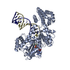
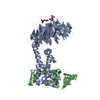
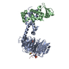
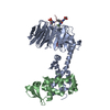
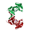
Mass: 63329.492 Da / Num. of mol.: 1 / Fragment: HELICASE DOMAIN, RESIDUES 242-794
Source method: isolated from a genetically manipulated source
Source: (gene. exp.) ANAS PLATYRHYNCHOS (mallard) / Plasmid: PFASTBAC / Cell line (production host): High Five / Production host: 
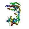
TRICHOPLUSIA NI (cabbage looper) / References: UniProt: D3TI84
Sequence detailsADDITIONAL GAM AT N-TERMINUS AFTER CLEAVAGE OF HIS-TAG

-
Experimental details

-
Experiment

ExperimentMethod: X-RAY DIFFRACTION / Number of used crystals: 1

-
Sample preparation

CrystalDensity Matthews: 2.75 Å3/Da / Density % sol: 55.5 % / Description: NONE
Crystal growDetails: 12 MG PER ML PROTEIN AND 0.1 M NACL, 0.1M M TRI-SODIUM CITRATE PH 5.6, 12% PEG 4000.

-
Data collection

DiffractionMean temperature: 100 K
Diffraction sourceSource: SYNCHROTRON / Site: ESRF / Beamline: ID14-4 / Wavelength: 0.976
DetectorType: ADSC CCD / Detector: CCD / Date: Mar 30, 2011
RadiationProtocol: SINGLE WAVELENGTH / Monochromatic (M) / Laue (L): M / Scattering type: x-ray
Radiation wavelengthWavelength: 0.976 Å / Relative weight: 1
ReflectionResolution: 3→50 Å / Num. obs: 13392 / % possible obs: 99.3 % / Observed criterion σ(I): 0 / Redundancy: 3.79 % / Rmerge(I) obs: 0.06 / Net I/σ(I): 16
Reflection shellResolution: 3→3.1 Å / Redundancy: 3.81 % / Rmerge(I) obs: 0.71 / Mean I/σ(I) obs: 1.8 / % possible all: 99.8

-
Processing

Software
NameVersionClassification
REFMAC5.6.0116refinement
XDSdata reduction
XSCALEdata scaling
SHELXphasing
RefinementMethod to determine structure: SAD
Starting model: NONE

Resolution: 3→33.59 Å / Cor.coef. Fo:Fc: 0.948 / Cor.coef. Fo:Fc free: 0.901 / SU B: 22.598 / SU ML: 0.41 / Cross valid method: THROUGHOUT / ESU R Free: 0.5 / Stereochemistry target values: MAXIMUM LIKELIHOOD
Details: HYDROGENS HAVE BEEN ADDED IN THE RIDING POSITIONS. U VALUES REFINED INDIVIDUALLY
RfactorNum. reflection% reflectionSelection details
Rfree0.29652 646 4.8 %RANDOM
Rwork0.21111 ---
obs0.21511 12746 99.3 %-
Solvent computationIon probe radii: 0.8 Å / Shrinkage radii: 0.8 Å / VDW probe radii: 1.2 Å / Solvent model: MASK
Displacement parametersBiso mean: 81.432 Å2
Baniso -1Baniso -2Baniso -3
1--3.54 Å2-1.77 Å20 Å2
2---3.54 Å20 Å2
3---5.31 Å2
Refinement stepCycle: LAST / Resolution: 3→33.59 Å
ProteinNucleic acidLigandSolventTotal
Num. atoms3852 0 0 0 3852
Refine LS restraints
Refine-IDTypeDev idealDev ideal targetNumber
X-RAY DIFFRACTIONr_bond_refined_d0.0050.023936
X-RAY DIFFRACTIONr_bond_other_d0.0010.022693
X-RAY DIFFRACTIONr_angle_refined_deg0.9561.9645307
X-RAY DIFFRACTIONr_angle_other_deg0.88936603
X-RAY DIFFRACTIONr_dihedral_angle_1_deg5.1665475
X-RAY DIFFRACTIONr_dihedral_angle_2_deg35.60924.973187
X-RAY DIFFRACTIONr_dihedral_angle_3_deg17.01315753
X-RAY DIFFRACTIONr_dihedral_angle_4_deg13.2851523
X-RAY DIFFRACTIONr_chiral_restr0.0950.2616
X-RAY DIFFRACTIONr_gen_planes_refined0.0030.024265
X-RAY DIFFRACTIONr_gen_planes_other0.0010.02742
X-RAY DIFFRACTIONr_nbd_refined
X-RAY DIFFRACTIONr_nbd_other
X-RAY DIFFRACTIONr_nbtor_refined
X-RAY DIFFRACTIONr_nbtor_other
X-RAY DIFFRACTIONr_xyhbond_nbd_refined
X-RAY DIFFRACTIONr_xyhbond_nbd_other
X-RAY DIFFRACTIONr_metal_ion_refined
X-RAY DIFFRACTIONr_metal_ion_other
X-RAY DIFFRACTIONr_symmetry_vdw_refined
X-RAY DIFFRACTIONr_symmetry_vdw_other
X-RAY DIFFRACTIONr_symmetry_hbond_refined
X-RAY DIFFRACTIONr_symmetry_hbond_other
X-RAY DIFFRACTIONr_symmetry_metal_ion_refined
X-RAY DIFFRACTIONr_symmetry_metal_ion_other
X-RAY DIFFRACTIONr_mcbond_it
X-RAY DIFFRACTIONr_mcbond_other
X-RAY DIFFRACTIONr_mcangle_it
X-RAY DIFFRACTIONr_mcangle_other
X-RAY DIFFRACTIONr_scbond_it
X-RAY DIFFRACTIONr_scbond_other
X-RAY DIFFRACTIONr_scangle_it
X-RAY DIFFRACTIONr_scangle_other
X-RAY DIFFRACTIONr_long_range_B_refined
X-RAY DIFFRACTIONr_long_range_B_other
X-RAY DIFFRACTIONr_rigid_bond_restr
X-RAY DIFFRACTIONr_sphericity_free
X-RAY DIFFRACTIONr_sphericity_bonded
LS refinement shellResolution: 3→3.078 Å / Total num. of bins used: 20
RfactorNum. reflection% reflection
Rfree0.348 59 -
Rwork0.342 927 -
obs--99.7 %

+
About Yorodumi

-
News

-
Feb 9, 2022. New format data for meta-information of EMDB entries

New format data for meta-information of EMDB entries

  • Version 3 of the EMDB header file is now the official format.
  • The previous official version 1.9 will be removed from the archive.

Related info.:EMDB header

External links:wwPDB to switch to version 3 of the EMDB data model

-
Aug 12, 2020. Covid-19 info

Covid-19 info

URL: https://pdbjlvh1.pdbj.org/emnavi/covid19.php

New page: Covid-19 featured information page in EM Navigator.

Related info.:Covid-19 info / Mar 5, 2020. Novel coronavirus structure data

+
Mar 5, 2020. Novel coronavirus structure data

Novel coronavirus structure data

Related info.:Yorodumi Speices / Aug 12, 2020. Covid-19 info

External links:COVID-19 featured content - PDBj / Molecule of the Month (242):Coronavirus Proteases

+
Jan 31, 2019. EMDB accession codes are about to change! (news from PDBe EMDB page)

EMDB accession codes are about to change! (news from PDBe EMDB page)

  • The allocation of 4 digits for EMDB accession codes will soon come to an end. Whilst these codes will remain in use, new EMDB accession codes will include an additional digit and will expand incrementally as the available range of codes is exhausted. The current 4-digit format prefixed with “EMD-” (i.e. EMD-XXXX) will advance to a 5-digit format (i.e. EMD-XXXXX), and so on. It is currently estimated that the 4-digit codes will be depleted around Spring 2019, at which point the 5-digit format will come into force.
  • The EM Navigator/Yorodumi systems omit the EMD- prefix.

Related info.:Q: What is EMD? / ID/Accession-code notation in Yorodumi/EM Navigator

External links:EMDB Accession Codes are Changing Soon! / Contact to PDBj

+
Jul 12, 2017. Major update of PDB

Major update of PDB

  • wwPDB released updated PDB data conforming to the new PDBx/mmCIF dictionary.
  • This is a major update changing the version number from 4 to 5, and with Remediation, in which all the entries are updated.
  • In this update, many items about electron microscopy experimental information are reorganized (e.g. em_software).
  • Now, EM Navigator and Yorodumi are based on the updated data.

External links:wwPDB Remediation / Enriched Model Files Conforming to OneDep Data Standards Now Available in the PDB FTP Archive

-
Yorodumi

Thousand views of thousand structures

  • Yorodumi is a browser for structure data from EMDB, PDB, SASBDB, etc.
  • This page is also the successor to EM Navigator detail page, and also detail information page/front-end page for Omokage search.
  • The word "yorodu" (or yorozu) is an old Japanese word meaning "ten thousand". "mi" (miru) is to see.

Related info.:EMDB / PDB / SASBDB / Comparison of 3 databanks / Yorodumi Search / Aug 31, 2016. New EM Navigator & Yorodumi / Yorodumi Papers / Jmol/JSmol / Function and homology information / Changes in new EM Navigator and Yorodumi

Read more