 Movie
Movie Controller

[English] 日本語
 Yorodumi
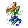
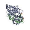
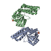
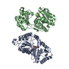
- PDB-3zxt: Dimeric structure of DAPK-1 catalytic domain in complex with AMPP... -
+ Open data

- Basic information
| Entry | Database: PDB / ID: 3zxt | ||||||
|---|---|---|---|---|---|---|---|
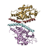
| Title | Dimeric structure of DAPK-1 catalytic domain in complex with AMPPCP- Mg | ||||||
 Components Components | DEATH-ASSOCIATED PROTEIN KINASE 1 | ||||||
 Keywords Keywords | TRANSFERASE / APOPTOSIS / ATP BINDING | ||||||
| Function / homology |  Function and homology information Function and homology informationcellular response to hydroperoxide / negative regulation of kinase activity / regulation of response to tumor cell / positive regulation of autophagic cell death / Caspase activation via Dependence Receptors in the absence of ligand / defense response to tumor cell / calcium/calmodulin-dependent protein kinase activity / regulation of NMDA receptor activity / syntaxin-1 binding / positive regulation of autophagy ...cellular response to hydroperoxide / negative regulation of kinase activity / regulation of response to tumor cell / positive regulation of autophagic cell death / Caspase activation via Dependence Receptors in the absence of ligand / defense response to tumor cell / calcium/calmodulin-dependent protein kinase activity / regulation of NMDA receptor activity / syntaxin-1 binding / positive regulation of autophagy / regulation of autophagy / apoptotic signaling pathway / cellular response to type II interferon / protein autophosphorylation / actin cytoskeleton / regulation of apoptotic process / protein phosphorylation / protein kinase activity / calmodulin binding / non-specific serine/threonine protein kinase / negative regulation of translation / postsynaptic density / intracellular signal transduction / positive regulation of apoptotic process / protein serine kinase activity / protein serine/threonine kinase activity / apoptotic process / negative regulation of apoptotic process / GTP binding / glutamatergic synapse / ATP binding / identical protein binding / nucleus / plasma membrane / cytoplasm Similarity search - Function | ||||||
| Biological species |  HOMO SAPIENS (human) HOMO SAPIENS (human) | ||||||
| Method |  X-RAY DIFFRACTION / SYNCHROTRON / MOLECULAR REPLACEMENT / Resolution: 2.65 Å X-RAY DIFFRACTION / SYNCHROTRON / MOLECULAR REPLACEMENT / Resolution: 2.65 Å | ||||||
 Authors Authors | de Diego, I. / Lehmann, F. / Wilmanns, M. | ||||||
 Citation Citation | Journal: To be Published Title: A Journey Through the Dap Kinase Architecture Authors: De Diego, I. / Kuper, J. / Lehmann, F. / Wilmanns, M. | ||||||
| History |
|
- Structure visualization
| Structure viewer | Molecule: MolmilJmol/JSmol |
|---|
- Downloads & links
-Download
| PDBx/mmCIF format | 3zxt.cif.gz | 401.5 KB | Display | PDBx/mmCIF format |
|---|---|---|---|---|
| PDB format | pdb3zxt.ent.gz | 328.9 KB | Display | PDB format |
| PDBx/mmJSON format | 3zxt.json.gz | Tree view | PDBx/mmJSON format | |
| Others |  Other downloads Other downloads |
-Validation report
| Arichive directory | https://data.pdbj.org/pub/pdb/validation_reports/zx/3zxtftp://data.pdbj.org/pub/pdb/validation_reports/zx/3zxt | HTTPS FTP |
|---|
-Related structure data
| Related structure data |  2y4pC  2y4vC  2w4jS S: Starting model for refinement C: citing same article ( |
|---|---|
| Similar structure data |


-Links
 PDBj
PDBj


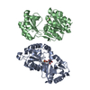
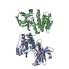
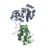
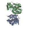
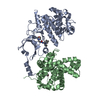
- Assembly
| Deposited unit | 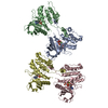
| ||||||||||||||||
|---|---|---|---|---|---|---|---|---|---|---|---|---|---|---|---|---|---|
| 1 | 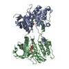
| ||||||||||||||||
| 2 | 
| ||||||||||||||||
| Unit cell |
| ||||||||||||||||
| Noncrystallographic symmetry (NCS) | NCS oper:
|
-Components
| #1: Protein | Mass: 32725.262 Da / Num. of mol.: 4 / Fragment: CATALYTIC DOMAIN, RESIDUES 1-285 Source method: isolated from a genetically manipulated source Source: (gene. exp.) HOMO SAPIENS (human) / Production host:  References: UniProt: P53355, non-specific serine/threonine protein kinase #2: Chemical | ChemComp-ACP /   Mass: 505.208 Da / Num. of mol.: 4 / Source method: obtained synthetically / Formula: C11H18N5O12P3 / Comment: AMP-PCP, energy-carrying molecule analogue*YM Mass: 505.208 Da / Num. of mol.: 4 / Source method: obtained synthetically / Formula: C11H18N5O12P3 / Comment: AMP-PCP, energy-carrying molecule analogue*YM#3: Chemical | ChemComp-MG /   Mass: 24.305 Da / Num. of mol.: 6 / Source method: obtained synthetically / Formula: Mg Mass: 24.305 Da / Num. of mol.: 6 / Source method: obtained synthetically / Formula: Mg#4: Water | ChemComp-HOH / |  Mass: 18.015 Da / Num. of mol.: 128 / Source method: isolated from a natural source / Formula: H2O Mass: 18.015 Da / Num. of mol.: 128 / Source method: isolated from a natural source / Formula: H2O |
|---|
-Experimental details
-Experiment
| Experiment | Method: X-RAY DIFFRACTION / Number of used crystals: 1 |
|---|
- Sample preparation
| Crystal | Density Matthews: 2.95 Å3/Da / Density % sol: 58.39 % / Description: NONE |
|---|---|
| Crystal grow | Details: 0.1 M HEPES PH7.0, 0.1M MGCL2, 15% PEG4000 |
-Data collection
| Diffraction | Mean temperature: 100 K |
|---|---|
| Diffraction source | Source: SYNCHROTRON / Site: ESRF  / Beamline: ID29 / Wavelength: 0.9235 / Beamline: ID29 / Wavelength: 0.9235 |
| Detector | Type: ADSC CCD / Detector: CCD / Date: Mar 5, 2011 / Details: MIRRORS |
| Radiation | Monochromator: SI / Protocol: SINGLE WAVELENGTH / Monochromatic (M) / Laue (L): M / Scattering type: x-ray |
| Radiation wavelength | Wavelength: 0.9235 Å / Relative weight: 1 |
| Reflection | Resolution: 2.65→45.71 Å / Num. obs: 39321 / % possible obs: 99.8 % / Observed criterion σ(I): 2.5 / Redundancy: 4 % / Rmerge(I) obs: 0.08 / Net I/σ(I): 10.2 |
| Reflection shell | Resolution: 2.65→2.79 Å / Redundancy: 4 % / Rmerge(I) obs: 0.47 / Mean I/σ(I) obs: 2.5 / % possible all: 97.4 |
- Processing
| Software |
| ||||||||||||||||||||||||||||||||||||||||||||||||||||||||||||||||||||||||||||||||||||||||||||||||||||||||||||||||||||||||||||||||||||||||||||||||||||||||||||||||||||||||||||||||||||||
|---|---|---|---|---|---|---|---|---|---|---|---|---|---|---|---|---|---|---|---|---|---|---|---|---|---|---|---|---|---|---|---|---|---|---|---|---|---|---|---|---|---|---|---|---|---|---|---|---|---|---|---|---|---|---|---|---|---|---|---|---|---|---|---|---|---|---|---|---|---|---|---|---|---|---|---|---|---|---|---|---|---|---|---|---|---|---|---|---|---|---|---|---|---|---|---|---|---|---|---|---|---|---|---|---|---|---|---|---|---|---|---|---|---|---|---|---|---|---|---|---|---|---|---|---|---|---|---|---|---|---|---|---|---|---|---|---|---|---|---|---|---|---|---|---|---|---|---|---|---|---|---|---|---|---|---|---|---|---|---|---|---|---|---|---|---|---|---|---|---|---|---|---|---|---|---|---|---|---|---|---|---|---|---|
| Refinement | Method to determine structure: MOLECULAR REPLACEMENT Starting model: PDB ENTRY 2W4J Resolution: 2.65→106.83 Å / Cor.coef. Fo:Fc: 0.937 / Cor.coef. Fo:Fc free: 0.893 / SU B: 29.35 / SU ML: 0.303 / Cross valid method: THROUGHOUT / ESU R: 0.694 / ESU R Free: 0.354 / Stereochemistry target values: MAXIMUM LIKELIHOOD Details: HYDROGENS HAVE BEEN USED IF PRESENT IN THE INPUT. U VALUES WITH TLS ADDED.
| ||||||||||||||||||||||||||||||||||||||||||||||||||||||||||||||||||||||||||||||||||||||||||||||||||||||||||||||||||||||||||||||||||||||||||||||||||||||||||||||||||||||||||||||||||||||
| Solvent computation | Ion probe radii: 0.8 Å / Shrinkage radii: 0.8 Å / VDW probe radii: 1.2 Å / Solvent model: MASK | ||||||||||||||||||||||||||||||||||||||||||||||||||||||||||||||||||||||||||||||||||||||||||||||||||||||||||||||||||||||||||||||||||||||||||||||||||||||||||||||||||||||||||||||||||||||
| Displacement parameters | Biso mean: 56.865 Å2
| ||||||||||||||||||||||||||||||||||||||||||||||||||||||||||||||||||||||||||||||||||||||||||||||||||||||||||||||||||||||||||||||||||||||||||||||||||||||||||||||||||||||||||||||||||||||
| Refinement step | Cycle: LAST / Resolution: 2.65→106.83 Å
| ||||||||||||||||||||||||||||||||||||||||||||||||||||||||||||||||||||||||||||||||||||||||||||||||||||||||||||||||||||||||||||||||||||||||||||||||||||||||||||||||||||||||||||||||||||||
| Refine LS restraints |
|