Protein / Protein/peptide , 2 types, 3 molecules ABQ
#1: Protein
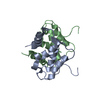
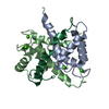
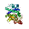
PROTEINS100-A4 / CALVASCULIN / METASTASIN / PLACENTAL CALCIUM-BINDING PROTEIN / PROTEIN MTS1 / S100 CALCIUM-BINDING ...CALVASCULIN / METASTASIN / PLACENTAL CALCIUM-BINDING PROTEIN / PROTEIN MTS1 / S100 CALCIUM-BINDING PROTEIN A4 / S100A4
Mass: 12018.653 Da / Num. of mol.: 2 / Mutation: YES Source method: isolated from a genetically manipulated source Source: (gene. exp.) HOMO SAPIENS (human) / Cell line: HEK / Plasmid: PBH4 / Production host: ESCHERICHIA COLI (E. coli) / Strain (production host): BL21(DE3) / References: UniProt: P26447
#2: Protein/peptide
MYOSIN-9 / CELLULAR MYOSIN HEAVY CHAIN\ / TYPE A / MYOSIN HEAVY CHAIN 9 / MYOSIN HEAVY CHAIN\ / NON-MUSCLE IIA ...CELLULAR MYOSIN HEAVY CHAIN\ / TYPE A / MYOSIN HEAVY CHAIN 9 / MYOSIN HEAVY CHAIN\ / NON-MUSCLE IIA / NON-MUSCLE MYOSIN HEAVY CHAIN A / NMMHC-A / NON-MUSCLE MYOSIN HEAVY CHAIN IIA / NMMHC II-A / NMMHC-IIA
Mass: 5358.213 Da / Num. of mol.: 1 / Fragment: RESIDUES 1893-1937 / Mutation: YES Source method: isolated from a genetically manipulated source Source: (gene. exp.) HOMO SAPIENS (human) / Cell line: HEK / Plasmid: PBH4 / Production host: ESCHERICHIA COLI (E. coli) / Strain (production host): BL21(DE3) / References: UniProt: P35579
Mass: 18.015 Da / Num. of mol.: 201 / Source method: isolated from a natural source / Formula: H2O
-
Details
Compound details
ENGINEERED RESIDUE IN CHAIN A, CYS 3 TO SER ENGINEERED RESIDUE IN CHAIN A, PHE 45 TO TRP ENGINEERED ...ENGINEERED RESIDUE IN CHAIN A, CYS 3 TO SER ENGINEERED RESIDUE IN CHAIN A, PHE 45 TO TRP ENGINEERED RESIDUE IN CHAIN A, CYS 81 TO SER ENGINEERED RESIDUE IN CHAIN A, CYS 86 TO SER ENGINEERED RESIDUE IN CHAIN B, CYS 3 TO SER ENGINEERED RESIDUE IN CHAIN B, PHE 45 TO TRP ENGINEERED RESIDUE IN CHAIN B, CYS 81 TO SER ENGINEERED RESIDUE IN CHAIN B, CYS 86 TO SER ENGINEERED RESIDUE IN CHAIN Q, ARG 1893 TO TYR
-
Experimental details
-
Experiment
Experiment
Method: X-RAY DIFFRACTION / Number of used crystals: 1
-
Sample preparation
Crystal
Density Matthews: 2.37 Å3/Da / Density % sol: 48.13 % / Description: NONE
Crystal grow
pH: 5.6 Details: 30% PEG 4000, 0.2 M NA-ACETATE PH 5.6, 0.1 M NA-CITRATE.
Resolution: 1.94→19.936 Å / SU ML: 0.2 / σ(F): 1.35 / Phase error: 17.55 / Stereochemistry target values: ML Details: CHAIN A RES MET-1 AND RES ASP95- -LYS101, CHAIN B RES LYS100-LYS101 AND CHAIN Q ARG1936- -LYS1937, COULD NOT BE MODELLED DUE TO MISSING ELECTRON DENSITY
Rfactor
Num. reflection
% reflection
Rfree
0.2064
1129
5.1 %
Rwork
0.1753
-
-
obs
0.1768
22109
99.93 %
Solvent computation
Shrinkage radii: 0.83 Å / VDW probe radii: 1.1 Å / Solvent model: FLAT BULK SOLVENT MODEL / Bsol: 34.329 Å2 / ksol: 0.341 e/Å3
Displacement parameters
Biso mean: 24.04 Å2
Baniso -1
Baniso -2
Baniso -3
1-
0.143 Å2
0 Å2
0 Å2
2-
-
0.143 Å2
0 Å2
3-
-
-
-0.2861 Å2
Refinement step
Cycle: LAST / Resolution: 1.94→19.936 Å
Protein
Nucleic acid
Ligand
Solvent
Total
Num. atoms
1907
0
18
201
2126
Refine LS restraints
Refine-ID
Type
Dev ideal
Number
X-RAY DIFFRACTION
f_bond_d
0.007
1966
X-RAY DIFFRACTION
f_angle_d
0.671
2615
X-RAY DIFFRACTION
f_dihedral_angle_d
12.497
758
X-RAY DIFFRACTION
f_chiral_restr
0.051
275
X-RAY DIFFRACTION
f_plane_restr
0.003
342
LS refinement shell
Resolution (Å)
Rfactor Rfree
Num. reflection Rfree
Rfactor Rwork
Num. reflection Rwork
Refine-ID
% reflection obs (%)
1.9401-2.0283
0.2581
126
0.2236
2561
X-RAY DIFFRACTION
100
2.0283-2.1351
0.2244
144
0.1895
2567
X-RAY DIFFRACTION
100
2.1351-2.2687
0.2147
166
0.1749
2541
X-RAY DIFFRACTION
100
2.2687-2.4436
0.2313
146
0.1723
2584
X-RAY DIFFRACTION
100
2.4436-2.689
0.2185
151
0.1805
2591
X-RAY DIFFRACTION
100
2.689-3.0768
0.2421
131
0.1819
2627
X-RAY DIFFRACTION
100
3.0768-3.8718
0.1681
145
0.1649
2660
X-RAY DIFFRACTION
100
3.8718-19.9374
0.1812
120
0.1647
2849
X-RAY DIFFRACTION
100
Refinement TLS params.
Method: refined / Refine-ID: X-RAY DIFFRACTION
ID
L11 (°2)
L12 (°2)
L13 (°2)
L22 (°2)
L23 (°2)
L33 (°2)
S11 (Å °)
S12 (Å °)
S13 (Å °)
S21 (Å °)
S22 (Å °)
S23 (Å °)
S31 (Å °)
S32 (Å °)
S33 (Å °)
T11 (Å2)
T12 (Å2)
T13 (Å2)
T22 (Å2)
T23 (Å2)
T33 (Å2)
Origin x (Å)
Origin y (Å)
Origin z (Å)
1
0.2816
0.4689
0.1058
1.0058
-0.2316
0.6905
-0.0219
-0.0677
0.0045
-0.0787
0.0374
0.0683
0.0518
-0.0162
0.0077
0.0661
0.0099
0.0007
0.0805
0.0243
0.0709
19.4054
2.3887
11.539
2
0.5451
-0.2854
0.0081
0.4651
-0.3475
0.9598
-0.0472
-0.2828
-0.0148
-0.0015
-0.1169
-0.2986
-0.0307
0.3666
-0.0404
0.0567
-0.0269
-0.0306
0.2408
0.0182
0.1546
39.5072
10.389
15.3482
3
0.2502
-0.0365
-0.2904
0.1478
-0.0509
0.2369
-0.0309
-0.0115
-0.1725
-0.0849
0.0544
-0.1001
0.2265
0.0504
-0.0061
0.1114
-0.0243
0.0304
0.0879
-0.0037
0.1021
27.8313
7.5467
-0.2418
4
0.0396
0.0356
-0.053
0.0421
0.0019
0.0469
0.0772
-0.0201
-0.1194
-0.0393
-0.0549
0.5315
0.0083
-0.2808
0
0.1194
0.009
0.0064
0.1278
0.0137
0.1394
16.023
-2.1429
24.528
5
0.0577
0.0934
-0.0089
0.1475
-0.0212
-0.002
-0.1159
-0.2805
0.0656
0.3524
0.0489
0.4245
-0.0017
0.0406
-0.0012
0.3143
0.0389
0.01
0.1747
-0.0037
0.1035
17.8175
11.7347
20.0613
6
0.1031
0.04
0.0085
-0.0018
-0.0143
0.0374
0.0835
0.0822
0.2437
0.0917
0.0312
-0.0738
-0.2732
0.0494
0.0001
0.1132
-0.0271
0.003
0.1309
0.0011
0.1304
30.4985
20.218
7.7556
7
0.2175
0.0818
0.2118
0.2136
0.2235
0.3346
-0.1337
0.0523
0.0307
-0.1821
-0.3508
-0.1961
-0.1848
0.1865
-0.0316
0.2282
-0.0314
0.0298
0.1329
0.0541
0.1532
36.1526
12.4065
-3.339
Refinement TLS group
ID
Refine-ID
Refine TLS-ID
Selection details
1
X-RAY DIFFRACTION
1
(CHAINAANDRESID2:94)
2
X-RAY DIFFRACTION
2
(CHAINBANDRESID1:74)
3
X-RAY DIFFRACTION
3
(CHAINBANDRESID75:99)
4
X-RAY DIFFRACTION
4
(CHAINQANDRESID1893:1902)
5
X-RAY DIFFRACTION
5
(CHAINQANDRESID1903:1908)
6
X-RAY DIFFRACTION
6
(CHAINQANDRESID1909:1930)
7
X-RAY DIFFRACTION
7
(CHAINQANDRESID1931:1935)
+
About Yorodumi
-
News
-
Feb 9, 2022. New format data for meta-information of EMDB entries
New format data for meta-information of EMDB entries
Version 3 of the EMDB header file is now the official format.
The previous official version 1.9 will be removed from the archive.
In the structure databanks used in Yorodumi, some data are registered as the other names, "COVID-19 virus" and "2019-nCoV". Here are the details of the virus and the list of structure data.
Jan 31, 2019. EMDB accession codes are about to change! (news from PDBe EMDB page)
EMDB accession codes are about to change! (news from PDBe EMDB page)
The allocation of 4 digits for EMDB accession codes will soon come to an end. Whilst these codes will remain in use, new EMDB accession codes will include an additional digit and will expand incrementally as the available range of codes is exhausted. The current 4-digit format prefixed with “EMD-” (i.e. EMD-XXXX) will advance to a 5-digit format (i.e. EMD-XXXXX), and so on. It is currently estimated that the 4-digit codes will be depleted around Spring 2019, at which point the 5-digit format will come into force.
The EM Navigator/Yorodumi systems omit the EMD- prefix.
Related info.:Q: What is EMD? / ID/Accession-code notation in Yorodumi/EM Navigator
Yorodumi is a browser for structure data from EMDB, PDB, SASBDB, etc.
This page is also the successor to EM Navigator detail page, and also detail information page/front-end page for Omokage search.
The word "yorodu" (or yorozu) is an old Japanese word meaning "ten thousand". "mi" (miru) is to see.
Related info.:EMDB / PDB / SASBDB / Comparison of 3 databanks / Yorodumi Search / Aug 31, 2016. New EM Navigator & Yorodumi / Yorodumi Papers / Jmol/JSmol / Function and homology information / Changes in new EM Navigator and Yorodumi
 Movie
Movie Controller

 Yorodumi
 Open data
 Basic information
 Components
 Keywords
 Function and homology information
 HOMO SAPIENS (human)
 X-RAY DIFFRACTION /
  Authors
 Citation
 Structure visualization
 Downloads & links
 Other downloads
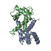


 PDBj
PDBj


 Assembly
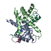


 Mass: 40.078 Da / Num. of mol.: 4 / Source method: obtained synthetically / Formula: Ca
 Mass: 59.044 Da / Num. of mol.: 2 / Source method: obtained synthetically / Formula: C2H3O2
 Mass: 42.020 Da / Num. of mol.: 2 / Source method: obtained synthetically / Formula: N3
 Sample preparation
 / Beamline: ID23-2 / Wavelength: 0.873
  Processing