 Movie
Movie Controller

[English] 日本語
 Yorodumi
- PDB-4cfr: Ca-bound S100A4 C3S, C81S, C86S and F45W mutant complexed with no... -
+ Open data

- Basic information
| Entry | Database: PDB / ID: 4cfr | |||||||||
|---|---|---|---|---|---|---|---|---|---|---|
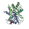
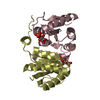
| Title | Ca-bound S100A4 C3S, C81S, C86S and F45W mutant complexed with non- muscle myosin IIA | |||||||||
 Components Components |
| |||||||||
 Keywords Keywords | CA-BINDING PROTEIN/MOTOR PROTEIN / CA-BINDING PROTEIN-MOTOR PROTEIN COMPLEX / CA-BINDING / EF-HAND | |||||||||
| Function / homology |  Function and homology information Function and homology informationnegative regulation of actin filament severing / uropod organization / regulation of plasma membrane repair / establishment of meiotic spindle localization / cytokinetic process / myosin II filament / cortical granule exocytosis / establishment of T cell polarity / blood vessel endothelial cell migration / actomyosin contractile ring ...negative regulation of actin filament severing / uropod organization / regulation of plasma membrane repair / establishment of meiotic spindle localization / cytokinetic process / myosin II filament / cortical granule exocytosis / establishment of T cell polarity / blood vessel endothelial cell migration / actomyosin contractile ring / cortical granule / positive regulation of protein processing in phagocytic vesicle / uropod / regulated exocytosis / cytoplasmic actin-based contraction involved in cell motility / RAGE receptor binding / meiotic spindle organization / lysosome localization / actin filament-based movement / actomyosin / myosin filament / myoblast fusion / plasma membrane repair / RHO GTPases Activate ROCKs / actomyosin structure organization / RHO GTPases activate CIT / Sema4D induced cell migration and growth-cone collapse / myosin II complex / platelet formation / Sensory processing of sound by outer hair cells of the cochlea / CD163 mediating an anti-inflammatory response / leukocyte migration / Sensory processing of sound by inner hair cells of the cochlea / phagocytosis, engulfment / EPHA-mediated growth cone collapse / microfilament motor activity / chemoattractant activity / endodermal cell differentiation / cell leading edge / membrane protein ectodomain proteolysis / cleavage furrow / RHO GTPases activate PAKs / cytoskeletal motor activity / brush border / transition metal ion binding / immunological synapse / epithelial to mesenchymal transition / monocyte differentiation / RHO GTPases activate PKNs / ruffle / stress fiber / protein-membrane adaptor activity / integrin-mediated signaling pathway / FCGR3A-mediated phagocytosis / Translocation of SLC2A4 (GLUT4) to the plasma membrane / adherens junction / neuromuscular junction / ADP binding / Regulation of actin dynamics for phagocytic cup formation / platelet aggregation / integrin binding / spindle / cytoplasmic side of plasma membrane / calcium-dependent protein binding / actin filament binding / Signaling by ALK fusions and activated point mutants / regulation of cell shape / protein transport / actin cytoskeleton / extracellular matrix / virus receptor activity / actin binding / actin cytoskeleton organization / angiogenesis / in utero embryonic development / positive regulation of canonical NF-kappaB signal transduction / calmodulin binding / nuclear body / cadherin binding / protein domain specific binding / focal adhesion / calcium ion binding / symbiont entry into host cell / perinuclear region of cytoplasm / cell surface / Golgi apparatus / protein homodimerization activity / protein-containing complex / : / RNA binding / extracellular exosome / extracellular region / nucleoplasm / ATP binding / membrane / identical protein binding / nucleus / plasma membrane / cytoplasm / cytosol Similarity search - Function | |||||||||
| Biological species |  HOMO SAPIENS (human) HOMO SAPIENS (human) | |||||||||
| Method |  X-RAY DIFFRACTION / SYNCHROTRON / MOLECULAR REPLACEMENT / Resolution: 1.4 Å X-RAY DIFFRACTION / SYNCHROTRON / MOLECULAR REPLACEMENT / Resolution: 1.4 Å | |||||||||
 Authors Authors | Duelli, A. / Kiss, B. / Lundholm, I. / Bodor, A. / Radnai, L. / Petoukhov, M. / Svergun, D. / Nyitray, L. / Katona, G. | |||||||||
 Citation Citation | Journal: Plos One / Year: 2014 Title: The C-Terminal Random Coil Region Tunes the Ca2+-Binding Affinity of S100A4 Through Conformational Activation. Authors: Duelli, A. / Kiss, B. / Lundholm, I. / Bodor, A. / Radnai, L. / Petoukhov, M. / Svergun, D. / Nyitray, L. / Katona, G. | |||||||||
| History |
|
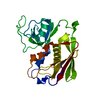
- Structure visualization
| Structure viewer | Molecule: MolmilJmol/JSmol |
|---|
- Downloads & links
-Download
| PDBx/mmCIF format | 4cfr.cif.gz | 164.8 KB | Display | PDBx/mmCIF format |
|---|---|---|---|---|
| PDB format | pdb4cfr.ent.gz | 132.3 KB | Display | PDB format |
| PDBx/mmJSON format | 4cfr.json.gz | Tree view | PDBx/mmJSON format | |
| Others |  Other downloads Other downloads |
-Validation report
| Arichive directory | https://data.pdbj.org/pub/pdb/validation_reports/cf/4cfrftp://data.pdbj.org/pub/pdb/validation_reports/cf/4cfr | HTTPS FTP |
|---|
-Related structure data
| Related structure data | 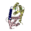 4cfqC  3zwhS C: citing same article ( S: Starting model for refinement |
|---|---|
| Similar structure data |
-Links
 PDBj
PDBj


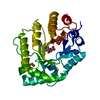
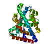
- Assembly
| Deposited unit | 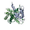
| ||||||||
|---|---|---|---|---|---|---|---|---|---|
| 1 |
| ||||||||
| Unit cell |
| ||||||||
| Components on special symmetry positions |
|
-Components
| #1: Protein | Mass: 12018.653 Da / Num. of mol.: 2 / Fragment: RESDIUES 1-101 / Mutation: YES Source method: isolated from a genetically manipulated source Source: (gene. exp.) HOMO SAPIENS (human) / Cell line: HEK / Plasmid: PBH4 / Production host:  #2: Protein/peptide | | Mass: 5358.213 Da / Num. of mol.: 1 / Fragment: RESIDUES 1893-1935 / Mutation: YES Source method: isolated from a genetically manipulated source Source: (gene. exp.) HOMO SAPIENS (human) / Cell line: HEK / Plasmid: PBH4 / Production host: #3: Chemical | ChemComp-CA /   Mass: 40.078 Da / Num. of mol.: 4 / Source method: obtained synthetically / Formula: Ca Mass: 40.078 Da / Num. of mol.: 4 / Source method: obtained synthetically / Formula: Ca#4: Water | ChemComp-HOH / |  Mass: 18.015 Da / Num. of mol.: 253 / Source method: isolated from a natural source / Formula: H2O Mass: 18.015 Da / Num. of mol.: 253 / Source method: isolated from a natural source / Formula: H2O |
|---|
-Experimental details
-Experiment
| Experiment | Method: X-RAY DIFFRACTION / Number of used crystals: 1 |
|---|
- Sample preparation
| Crystal | Density Matthews: 2.42 Å3/Da / Density % sol: 49.31 % / Description: NONE |
|---|---|
| Crystal grow | pH: 4.6 Details: 0.2 M AMMONIUM ACETATE, 0.1M SODIUM ACETATE PH4.6, 30% W/V PEG 4000 |
-Data collection
| Diffraction | Mean temperature: 100 K |
|---|---|
| Diffraction source | Source: SYNCHROTRON / Site: ESRF  / Beamline: ID23-1 / Wavelength: 1.0722 / Beamline: ID23-1 / Wavelength: 1.0722 |
| Detector | Type: ADSC CCD / Detector: CCD / Date: Sep 11, 2011 |
| Radiation | Monochromator: SI(111) / Protocol: SINGLE WAVELENGTH / Monochromatic (M) / Laue (L): M / Scattering type: x-ray |
| Radiation wavelength | Wavelength: 1.0722 Å / Relative weight: 1 |
| Reflection | Resolution: 1.4→29.1 Å / Num. obs: 57504 / % possible obs: 99.5 % / Observed criterion σ(I): 3 / Redundancy: 9 % / Biso Wilson estimate: 16 Å2 / Rmerge(I) obs: 0.06 / Net I/σ(I): 18.6 |
| Reflection shell | Resolution: 1.4→1.48 Å / Redundancy: 6.4 % / Rmerge(I) obs: 0.76 / Mean I/σ(I) obs: 2.1 / % possible all: 99.1 |
- Processing
| Software |
| ||||||||||||||||||||||||||||||||||||||||||||||||||||||||||||||||||||||||||||||||||||||||||||||||||
|---|---|---|---|---|---|---|---|---|---|---|---|---|---|---|---|---|---|---|---|---|---|---|---|---|---|---|---|---|---|---|---|---|---|---|---|---|---|---|---|---|---|---|---|---|---|---|---|---|---|---|---|---|---|---|---|---|---|---|---|---|---|---|---|---|---|---|---|---|---|---|---|---|---|---|---|---|---|---|---|---|---|---|---|---|---|---|---|---|---|---|---|---|---|---|---|---|---|---|---|
| Refinement | Method to determine structure: MOLECULAR REPLACEMENT Starting model: PDB ENTRY 3ZWH Resolution: 1.4→29.09 Å / SU ML: 0.16 / σ(F): 1.99 / Phase error: 15.33 / Stereochemistry target values: ML
| ||||||||||||||||||||||||||||||||||||||||||||||||||||||||||||||||||||||||||||||||||||||||||||||||||
| Solvent computation | Shrinkage radii: 0.86 Å / VDW probe radii: 1.1 Å / Solvent model: FLAT BULK SOLVENT MODEL / Bsol: 45.229 Å2 / ksol: 0.407 e/Å3 | ||||||||||||||||||||||||||||||||||||||||||||||||||||||||||||||||||||||||||||||||||||||||||||||||||
| Displacement parameters | Biso mean: 25.4 Å2
| ||||||||||||||||||||||||||||||||||||||||||||||||||||||||||||||||||||||||||||||||||||||||||||||||||
| Refinement step | Cycle: LAST / Resolution: 1.4→29.09 Å
| ||||||||||||||||||||||||||||||||||||||||||||||||||||||||||||||||||||||||||||||||||||||||||||||||||
| Refine LS restraints |
| ||||||||||||||||||||||||||||||||||||||||||||||||||||||||||||||||||||||||||||||||||||||||||||||||||
| LS refinement shell |
|
