 Movie
Movie Controller

[English] 日本語
 Yorodumi
- PDB-2vvp: Crystal structure of Mycobacterium tuberculosis ribose-5-phosphat... -
+ Open data

- Basic information
| Entry | Database: PDB / ID: 2vvp | |||||||||
|---|---|---|---|---|---|---|---|---|---|---|
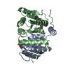
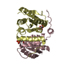
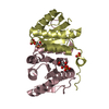
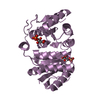
| Title | Crystal structure of Mycobacterium tuberculosis ribose-5-phosphate isomerase B in complex with its substrates ribose 5-phosphate and ribulose 5-phosphate | |||||||||
 Components Components | RIBOSE-5-PHOSPHATE ISOMERASE B | |||||||||
 Keywords Keywords | ISOMERASE / RPIB / RV2465C / RARE SUGAR / CARBOHYDRATE METABOLISM / PENTOSE PHOSPHATE PATHWAY | |||||||||
| Function / homology |  Function and homology information Function and homology informationD-allose catabolic process / ribose-5-phosphate isomerase / ribose-5-phosphate isomerase activity / pentose-phosphate shunt, non-oxidative branch / carbohydrate metabolic process / extracellular region Similarity search - Function | |||||||||
| Biological species |   MYCOBACTERIUM TUBERCULOSIS (bacteria) MYCOBACTERIUM TUBERCULOSIS (bacteria) | |||||||||
| Method |  X-RAY DIFFRACTION / SYNCHROTRON / MOLECULAR REPLACEMENT / Resolution: 1.65 Å X-RAY DIFFRACTION / SYNCHROTRON / MOLECULAR REPLACEMENT / Resolution: 1.65 Å | |||||||||
 Authors Authors | Kowalinski, E. / Roos, A.K. / Mariano, S. / Salmon, L. / Mowbray, S.L. | |||||||||
 Citation Citation | Journal: J.Mol.Biol. / Year: 2008 Title: D-Ribose-5-Phosphate Isomerase B from Escherichia Coli is Also a Functional D-Allose-6-Phosphate Isomerase, While the Mycobacterium Tuberculosis Enzyme is not. Authors: Roos, A.K. / Mariano, S. / Kowalinski, E. / Salmon, L. / Mowbray, S.L. | |||||||||
| History |
|
- Structure visualization
| Structure viewer | Molecule: MolmilJmol/JSmol |
|---|
- Downloads & links
-Download
| PDBx/mmCIF format | 2vvp.cif.gz | 179.4 KB | Display | PDBx/mmCIF format |
|---|---|---|---|---|
| PDB format | pdb2vvp.ent.gz | 144.5 KB | Display | PDB format |
| PDBx/mmJSON format | 2vvp.json.gz | Tree view | PDBx/mmJSON format | |
| Others |  Other downloads Other downloads |
-Validation report
| Arichive directory | https://data.pdbj.org/pub/pdb/validation_reports/vv/2vvpftp://data.pdbj.org/pub/pdb/validation_reports/vv/2vvp | HTTPS FTP |
|---|
-Related structure data
| Related structure data | 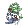 2vvoC  2vvqC 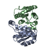 2vvrC  1uslS C: citing same article ( S: Starting model for refinement |
|---|---|
| Similar structure data |


-Links
 PDBj
PDBj- Assembly
| Deposited unit | 
| ||||||||||||||||||||
|---|---|---|---|---|---|---|---|---|---|---|---|---|---|---|---|---|---|---|---|---|---|
| 1 | 
| ||||||||||||||||||||
| 2 | 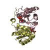
| ||||||||||||||||||||
| 3 | 
| ||||||||||||||||||||
| Unit cell |
| ||||||||||||||||||||
| Components on special symmetry positions |
| ||||||||||||||||||||
| Noncrystallographic symmetry (NCS) | NCS oper:
|
-Components
| #1: Protein | Mass: 17299.514 Da / Num. of mol.: 5 Source method: isolated from a genetically manipulated source Source: (gene. exp.) MYCOBACTERIUM TUBERCULOSIS (bacteria) / Strain: H37RV / Plasmid: PCRT7 / Production host: References: UniProt: Q79FD7, UniProt: P9WKD7*PLUS, ribose-5-phosphate isomerase #2: Sugar | ChemComp-R5P /   Type: saccharide / Mass: 230.110 Da / Num. of mol.: 5 / Source method: obtained synthetically / Formula: C5H11O8P Type: saccharide / Mass: 230.110 Da / Num. of mol.: 5 / Source method: obtained synthetically / Formula: C5H11O8P#3: Sugar | ChemComp-5RP /   Type: saccharide / Mass: 230.110 Da / Num. of mol.: 5 Type: saccharide / Mass: 230.110 Da / Num. of mol.: 5Source method: isolated from a genetically manipulated source Formula: C5H11O8P #4: Water | ChemComp-HOH / |  Mass: 18.015 Da / Num. of mol.: 981 / Source method: isolated from a natural source / Formula: H2O Mass: 18.015 Da / Num. of mol.: 981 / Source method: isolated from a natural source / Formula: H2OHas protein modification | N | |
|---|
-Experimental details
-Experiment
| Experiment | Method: X-RAY DIFFRACTION / Number of used crystals: 1 |
|---|
- Sample preparation
| Crystal | Density Matthews: 2.83 Å3/Da / Density % sol: 56.5 % / Description: NONE |
|---|---|
| Crystal grow | Details: 20% PEG 3000, 0.1 M TRIS PH 7, 0.2 M CA ACETATE |
-Data collection
| Diffraction | Mean temperature: 110 K |
|---|---|
| Diffraction source | Source: SYNCHROTRON / Site: ESRF  / Beamline: ID14-3 / Wavelength: 0.931 / Beamline: ID14-3 / Wavelength: 0.931 |
| Detector | Type: ADSC CCD / Detector: CCD / Date: May 5, 2005 |
| Radiation | Protocol: SINGLE WAVELENGTH / Monochromatic (M) / Laue (L): M / Scattering type: x-ray |
| Radiation wavelength | Wavelength: 0.931 Å / Relative weight: 1 |
| Reflection | Resolution: 1.65→35 Å / Num. obs: 115358 / % possible obs: 99.9 % / Observed criterion σ(I): 3 / Redundancy: 3.6 % / Biso Wilson estimate: 19.8 Å2 / Rmerge(I) obs: 0.07 / Net I/σ(I): 13.3 |
| Reflection shell | Resolution: 1.65→1.74 Å / Redundancy: 2.9 % / Rmerge(I) obs: 0.29 / Mean I/σ(I) obs: 3 / % possible all: 99.9 |
- Processing
| Software |
| ||||||||||||||||||||||||||||||||||||||||||||||||||||||||||||||||||||||||||||||||||||||||||||||||||||||||||||||||||||||||||||||||||||||||||||||||||||||||||||||||||||||||||||||||||||||
|---|---|---|---|---|---|---|---|---|---|---|---|---|---|---|---|---|---|---|---|---|---|---|---|---|---|---|---|---|---|---|---|---|---|---|---|---|---|---|---|---|---|---|---|---|---|---|---|---|---|---|---|---|---|---|---|---|---|---|---|---|---|---|---|---|---|---|---|---|---|---|---|---|---|---|---|---|---|---|---|---|---|---|---|---|---|---|---|---|---|---|---|---|---|---|---|---|---|---|---|---|---|---|---|---|---|---|---|---|---|---|---|---|---|---|---|---|---|---|---|---|---|---|---|---|---|---|---|---|---|---|---|---|---|---|---|---|---|---|---|---|---|---|---|---|---|---|---|---|---|---|---|---|---|---|---|---|---|---|---|---|---|---|---|---|---|---|---|---|---|---|---|---|---|---|---|---|---|---|---|---|---|---|---|
| Refinement | Method to determine structure: MOLECULAR REPLACEMENT Starting model: PDB ENTRY 1USL Resolution: 1.65→34.71 Å / Cor.coef. Fo:Fc: 0.963 / Cor.coef. Fo:Fc free: 0.955 / SU B: 1.501 / SU ML: 0.052 / Cross valid method: THROUGHOUT / ESU R: 0.082 / ESU R Free: 0.08 / Stereochemistry target values: MAXIMUM LIKELIHOOD / Details: HYDROGENS HAVE BEEN ADDED IN THE RIDING POSITIONS.
| ||||||||||||||||||||||||||||||||||||||||||||||||||||||||||||||||||||||||||||||||||||||||||||||||||||||||||||||||||||||||||||||||||||||||||||||||||||||||||||||||||||||||||||||||||||||
| Solvent computation | Ion probe radii: 0.8 Å / Shrinkage radii: 0.8 Å / VDW probe radii: 1.2 Å / Solvent model: MASK | ||||||||||||||||||||||||||||||||||||||||||||||||||||||||||||||||||||||||||||||||||||||||||||||||||||||||||||||||||||||||||||||||||||||||||||||||||||||||||||||||||||||||||||||||||||||
| Displacement parameters | Biso mean: 13.95 Å2
| ||||||||||||||||||||||||||||||||||||||||||||||||||||||||||||||||||||||||||||||||||||||||||||||||||||||||||||||||||||||||||||||||||||||||||||||||||||||||||||||||||||||||||||||||||||||
| Refinement step | Cycle: LAST / Resolution: 1.65→34.71 Å
| ||||||||||||||||||||||||||||||||||||||||||||||||||||||||||||||||||||||||||||||||||||||||||||||||||||||||||||||||||||||||||||||||||||||||||||||||||||||||||||||||||||||||||||||||||||||
| Refine LS restraints |
|