 Movie
Movie Controller

[English] 日本語
 Yorodumi
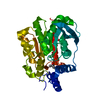
- PDB-1usl: Structure Of Mycobacterium tuberculosis Ribose-5-Phosphate Isomer... -
+ Open data

- Basic information
| Entry | Database: PDB / ID: 1usl | ||||||
|---|---|---|---|---|---|---|---|
| Title | Structure Of Mycobacterium tuberculosis Ribose-5-Phosphate Isomerase, RpiB, Rv2465c, Complexed With Phosphate. | ||||||
 Components Components | RIBOSE 5-PHOSPHATE ISOMERASE B | ||||||
 Keywords Keywords | ISOMERASE / PHOSPHOPENTOSE ISOMERASE B / PENTOSE PHOSPHATE PATHWAY / RV2465C / PHOSPHORIBOISOMERASE B / STRUCTURAL PROTEOMICS IN EUROPE / SPINE / STRUCTURAL GENOMICS | ||||||
| Function / homology |  Function and homology information Function and homology informationD-allose catabolic process / ribose-5-phosphate isomerase / ribose-5-phosphate isomerase activity / pentose-phosphate shunt, non-oxidative branch / extracellular region Similarity search - Function | ||||||
| Biological species |   MYCOBACTERIUM TUBERCULOSIS (bacteria) MYCOBACTERIUM TUBERCULOSIS (bacteria) | ||||||
| Method |  X-RAY DIFFRACTION / SYNCHROTRON / MOLECULAR REPLACEMENT / Resolution: 1.88 Å X-RAY DIFFRACTION / SYNCHROTRON / MOLECULAR REPLACEMENT / Resolution: 1.88 Å | ||||||
 Authors Authors | Roos, A.K. / Andersson, C.E. / Unge, T. / Jones, T.A. / Mowbray, S.L. | ||||||
 Citation Citation | Journal: J.Mol.Biol. / Year: 2004 Title: Mycobacterium Tuberculosis Ribose-5-Phosphate Isomerase Has a Known Fold, But a Novel Active Site Authors: Roos, A.K. / Andersson, C.E. / Bergfors, T. / Jacobsson, M. / Karlen, A. / Unge, T. / Jones, T.A. / Mowbray, S.L. | ||||||
| History |
| ||||||
| Remark 650 | HELIX DETERMINATION METHOD: AUTHOR PROVIDED. | ||||||
| Remark 700 | SHEET DETERMINATION METHOD: AUTHOR PROVIDED. |
- Structure visualization
| Structure viewer | Molecule: MolmilJmol/JSmol |
|---|
- Downloads & links
-Download
| PDBx/mmCIF format | 1usl.cif.gz | 166.4 KB | Display | PDBx/mmCIF format |
|---|---|---|---|---|
| PDB format | pdb1usl.ent.gz | 132.4 KB | Display | PDB format |
| PDBx/mmJSON format | 1usl.json.gz | Tree view | PDBx/mmJSON format | |
| Others |  Other downloads Other downloads |
-Validation report
| Arichive directory | https://data.pdbj.org/pub/pdb/validation_reports/us/1uslftp://data.pdbj.org/pub/pdb/validation_reports/us/1usl | HTTPS FTP |
|---|
-Related structure data
| Related structure data |  1nn4S S: Starting model for refinement |
|---|---|
| Similar structure data |


-Links
 PDBj
PDBj- Assembly
| Deposited unit | 
| ||||||||||||||||||||
|---|---|---|---|---|---|---|---|---|---|---|---|---|---|---|---|---|---|---|---|---|---|
| 1 | 
| ||||||||||||||||||||
| 2 | 
| ||||||||||||||||||||
| 3 | 
| ||||||||||||||||||||
| Unit cell |
| ||||||||||||||||||||
| Components on special symmetry positions |
| ||||||||||||||||||||
| Noncrystallographic symmetry (NCS) | NCS oper:
|
-Components
| #1: Protein | Mass: 18330.672 Da / Num. of mol.: 5 Source method: isolated from a genetically manipulated source Source: (gene. exp.) MYCOBACTERIUM TUBERCULOSIS (bacteria) / Strain: H37RV / Plasmid: PCR_T7 / Production host: References: UniProt: O53192, UniProt: P9WKD7*PLUS, ribose-5-phosphate isomerase #2: Chemical | ChemComp-PO4 /   Mass: 94.971 Da / Num. of mol.: 5 / Source method: obtained synthetically / Formula: PO4 Mass: 94.971 Da / Num. of mol.: 5 / Source method: obtained synthetically / Formula: PO4#3: Water | ChemComp-HOH / |  Mass: 18.015 Da / Num. of mol.: 579 / Source method: isolated from a natural source / Formula: H2O Mass: 18.015 Da / Num. of mol.: 579 / Source method: isolated from a natural source / Formula: H2OSequence details | N-TERMINAL 6-HIS TAG | |
|---|
-Experimental details
-Experiment
| Experiment | Method: X-RAY DIFFRACTION / Number of used crystals: 1 |
|---|
- Sample preparation
| Crystal | Density Matthews: 2.8 Å3/Da / Density % sol: 55.7 % | ||||||||||||||||||||||||||||||||||||||||||||||||||||||||
|---|---|---|---|---|---|---|---|---|---|---|---|---|---|---|---|---|---|---|---|---|---|---|---|---|---|---|---|---|---|---|---|---|---|---|---|---|---|---|---|---|---|---|---|---|---|---|---|---|---|---|---|---|---|---|---|---|---|
| Crystal grow | pH: 7.5 / Details: 1.26 M NAKPO4 PH 7.5 | ||||||||||||||||||||||||||||||||||||||||||||||||||||||||
| Crystal grow | *PLUS Temperature: 20 ℃ / pH: 6 / Method: vapor diffusion, sitting drop | ||||||||||||||||||||||||||||||||||||||||||||||||||||||||
| Components of the solutions | *PLUS
|
-Data collection
| Diffraction | Mean temperature: 110 K |
|---|---|
| Diffraction source | Source: SYNCHROTRON / Site: MAX II  / Beamline: I711 / Wavelength: 1.098 / Beamline: I711 / Wavelength: 1.098 |
| Detector | Type: MARRESEARCH / Detector: CCD / Date: Nov 15, 2002 |
| Radiation | Monochromator: SI(111) / Protocol: SINGLE WAVELENGTH / Monochromatic (M) / Laue (L): M / Scattering type: x-ray |
| Radiation wavelength | Wavelength: 1.098 Å / Relative weight: 1 |
| Reflection | Resolution: 1.88→81.65 Å / Num. obs: 73429 / % possible obs: 95.6 % / Observed criterion σ(I): 2 / Redundancy: 4.9 % / Rmerge(I) obs: 0.069 / Net I/σ(I): 9.6 |
| Reflection shell | Resolution: 1.88→1.98 Å / Redundancy: 4.8 % / Rmerge(I) obs: 0.335 / Mean I/σ(I) obs: 2.5 / % possible all: 90.6 |
| Reflection | *PLUS Highest resolution: 1.88 Å / Lowest resolution: 81.65 Å / Redundancy: 4.9 % / Num. measured all: 358802 / Rmerge(I) obs: 0.069 |
| Reflection shell | *PLUS % possible obs: 90.6 % / Redundancy: 4.8 % / Rmerge(I) obs: 0.335 / Mean I/σ(I) obs: 2.5 |
- Processing
| Software |
| ||||||||||||||||||||
|---|---|---|---|---|---|---|---|---|---|---|---|---|---|---|---|---|---|---|---|---|---|
| Refinement | Method to determine structure: MOLECULAR REPLACEMENT Starting model: PDB ENTRY 1NN4 Resolution: 1.88→81.65 Å / SU B: 2.581 / SU ML: 0.077 / Cross valid method: THROUGHOUT / ESU R: 0.127 / ESU R Free: 0.12 / Details: HYDROGENS HAVE BEEN ADDED IN THE RIDING POSITIONS
| ||||||||||||||||||||
| Displacement parameters | Biso mean: 20.846 Å2
| ||||||||||||||||||||
| Refinement step | Cycle: LAST / Resolution: 1.88→81.65 Å
| ||||||||||||||||||||
| Refinement | *PLUS Rfactor Rfree: 0.202 / Rfactor Rwork: 0.168 | ||||||||||||||||||||
| Solvent computation | *PLUS | ||||||||||||||||||||
| Displacement parameters | *PLUS | ||||||||||||||||||||
| Refine LS restraints | *PLUS
|
