 Movie
Movie Controller

[English] 日本語
 Yorodumi
- PDB-3znz: Crystal structure of OTULIN OTU domain (C129A) in complex with Me... -
+ Open data

- Basic information
| Entry | Database: PDB / ID: 3znz | ||||||
|---|---|---|---|---|---|---|---|
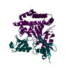
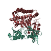
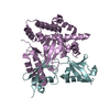
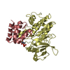
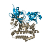
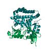
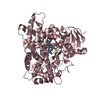
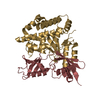
| Title | Crystal structure of OTULIN OTU domain (C129A) in complex with Met1- di ubiquitin | ||||||
 Components Components |
| ||||||
 Keywords Keywords | HYDROLASE | ||||||
| Function / homology |  Function and homology information Function and homology informationprotein linear deubiquitination / LUBAC complex / nucleotide-binding oligomerization domain containing 2 signaling pathway / regulation of tumor necrosis factor-mediated signaling pathway / sprouting angiogenesis / regulation of canonical Wnt signaling pathway / : / cysteine-type peptidase activity / Maturation of protein E / Maturation of protein E ...protein linear deubiquitination / LUBAC complex / nucleotide-binding oligomerization domain containing 2 signaling pathway / regulation of tumor necrosis factor-mediated signaling pathway / sprouting angiogenesis / regulation of canonical Wnt signaling pathway / : / cysteine-type peptidase activity / Maturation of protein E / Maturation of protein E / ER Quality Control Compartment (ERQC) / Myoclonic epilepsy of Lafora / FLT3 signaling by CBL mutants / IRAK2 mediated activation of TAK1 complex / Alpha-protein kinase 1 signaling pathway / Glycogen synthesis / IRAK1 recruits IKK complex / IRAK1 recruits IKK complex upon TLR7/8 or 9 stimulation / Prevention of phagosomal-lysosomal fusion / Endosomal Sorting Complex Required For Transport (ESCRT) / Membrane binding and targetting of GAG proteins / Negative regulation of FLT3 / Regulation of TBK1, IKKε (IKBKE)-mediated activation of IRF3, IRF7 / PTK6 Regulates RTKs and Their Effectors AKT1 and DOK1 / Regulation of TBK1, IKKε-mediated activation of IRF3, IRF7 upon TLR3 ligation / IRAK2 mediated activation of TAK1 complex upon TLR7/8 or 9 stimulation / Constitutive Signaling by NOTCH1 HD Domain Mutants / NOTCH2 Activation and Transmission of Signal to the Nucleus / TICAM1,TRAF6-dependent induction of TAK1 complex / TICAM1-dependent activation of IRF3/IRF7 / APC/C:Cdc20 mediated degradation of Cyclin B / Downregulation of ERBB4 signaling / APC-Cdc20 mediated degradation of Nek2A / Regulation of FZD by ubiquitination / p75NTR recruits signalling complexes / InlA-mediated entry of Listeria monocytogenes into host cells / TRAF6 mediated IRF7 activation in TLR7/8 or 9 signaling / NF-kB is activated and signals survival / TRAF6-mediated induction of TAK1 complex within TLR4 complex / Regulation of pyruvate metabolism / Pexophagy / Downregulation of ERBB2:ERBB3 signaling / Regulation of innate immune responses to cytosolic DNA / NRIF signals cell death from the nucleus / Regulation of PTEN localization / VLDLR internalisation and degradation / Activated NOTCH1 Transmits Signal to the Nucleus / Synthesis of active ubiquitin: roles of E1 and E2 enzymes / Translesion synthesis by REV1 / TICAM1, RIP1-mediated IKK complex recruitment / Regulation of BACH1 activity / Translesion synthesis by POLK / InlB-mediated entry of Listeria monocytogenes into host cell / JNK (c-Jun kinases) phosphorylation and activation mediated by activated human TAK1 / MAP3K8 (TPL2)-dependent MAPK1/3 activation / Activation of IRF3, IRF7 mediated by TBK1, IKKε (IKBKE) / Downregulation of TGF-beta receptor signaling / Translesion synthesis by POLI / Josephin domain DUBs / Gap-filling DNA repair synthesis and ligation in GG-NER / IKK complex recruitment mediated by RIP1 / PINK1-PRKN Mediated Mitophagy / TGF-beta receptor signaling in EMT (epithelial to mesenchymal transition) / Regulation of activated PAK-2p34 by proteasome mediated degradation / TNFR1-induced NF-kappa-B signaling pathway / TCF dependent signaling in response to WNT / Regulation of NF-kappa B signaling / activated TAK1 mediates p38 MAPK activation / Autodegradation of Cdh1 by Cdh1:APC/C / APC/C:Cdc20 mediated degradation of Securin / N-glycan trimming in the ER and Calnexin/Calreticulin cycle / Asymmetric localization of PCP proteins / NOTCH3 Activation and Transmission of Signal to the Nucleus / Regulation of signaling by CBL / Negative regulators of DDX58/IFIH1 signaling / Ubiquitin-dependent degradation of Cyclin D / Fanconi Anemia Pathway / Negative regulation of FGFR3 signaling / Peroxisomal protein import / SCF-beta-TrCP mediated degradation of Emi1 / Deactivation of the beta-catenin transactivating complex / NIK-->noncanonical NF-kB signaling / AUF1 (hnRNP D0) binds and destabilizes mRNA / TNFR2 non-canonical NF-kB pathway / Stabilization of p53 / Negative regulation of FGFR2 signaling / Negative regulation of FGFR4 signaling / Assembly of the pre-replicative complex / Vpu mediated degradation of CD4 / Downregulation of SMAD2/3:SMAD4 transcriptional activity / Negative regulation of FGFR1 signaling / Termination of translesion DNA synthesis / Cdc20:Phospho-APC/C mediated degradation of Cyclin A / EGFR downregulation / Regulation of TNFR1 signaling / Dectin-1 mediated noncanonical NF-kB signaling / Assembly Of The HIV Virion / Degradation of DVL / SMAD2/SMAD3:SMAD4 heterotrimer regulates transcription / Degradation of AXIN Similarity search - Function | ||||||
| Biological species |  HOMO SAPIENS (human) HOMO SAPIENS (human) | ||||||
| Method |  X-RAY DIFFRACTION / SYNCHROTRON / MOLECULAR REPLACEMENT / Resolution: 1.9 Å X-RAY DIFFRACTION / SYNCHROTRON / MOLECULAR REPLACEMENT / Resolution: 1.9 Å | ||||||
 Authors Authors | Keusekotten, K. / Elliott, P.R. / Glockner, L. / Kulathu, Y. / Wauer, T. / Krappmann, D. / Hofmann, K. / Komander, D. | ||||||
 Citation Citation | Journal: Cell(Cambridge,Mass.) / Year: 2013 Title: Otulin Antagonizes Lubac Signaling by Specifically Hydrolyzing met1-Linked Polyubiquitin. Authors: Keusekotten, K. / Elliott, P.R. / Glockner, L. / Fiil, B.K. / Damgaard, R.B. / Kulathu, Y. / Wauer, T. / Hospenthal, M.K. / Gyrd-Hansen, M. / Krappmann, D. / Hofmann, K. / Komander, D. | ||||||
| History |
|
- Structure visualization
| Structure viewer | Molecule: MolmilJmol/JSmol |
|---|
- Downloads & links
-Download
| PDBx/mmCIF format | 3znz.cif.gz | 107.7 KB | Display | PDBx/mmCIF format |
|---|---|---|---|---|
| PDB format | pdb3znz.ent.gz | 82 KB | Display | PDB format |
| PDBx/mmJSON format | 3znz.json.gz | Tree view | PDBx/mmJSON format | |
| Others |  Other downloads Other downloads |
-Validation report
| Arichive directory | https://data.pdbj.org/pub/pdb/validation_reports/zn/3znzftp://data.pdbj.org/pub/pdb/validation_reports/zn/3znz | HTTPS FTP |
|---|
-Related structure data
| Related structure data |  3znvSC  3znxC S: Starting model for refinement C: citing same article ( |
|---|---|
| Similar structure data |
-Links
 PDBj
PDBj


- Assembly
| Deposited unit | 
| ||||||||||||
|---|---|---|---|---|---|---|---|---|---|---|---|---|---|
| 1 |
| ||||||||||||
| Unit cell |
| ||||||||||||
| Components on special symmetry positions |
|
-Components
| #1: Protein | Mass: 31894.590 Da / Num. of mol.: 1 / Fragment: OTU DOMAIN, RESIDUES 80-352 / Mutation: YES Source method: isolated from a genetically manipulated source Source: (gene. exp.) HOMO SAPIENS (human) / Production host:  | ||||
|---|---|---|---|---|---|
| #2: Protein | Mass: 17135.654 Da / Num. of mol.: 1 Source method: isolated from a genetically manipulated source Source: (gene. exp.) HOMO SAPIENS (human) / Production host: | ||||
| #3: Chemical | ChemComp-SO4 /   Mass: 96.063 Da / Num. of mol.: 7 / Source method: obtained synthetically / Formula: SO4 Mass: 96.063 Da / Num. of mol.: 7 / Source method: obtained synthetically / Formula: SO4#4: Water | ChemComp-HOH / |  Mass: 18.015 Da / Num. of mol.: 370 / Source method: isolated from a natural source / Formula: H2O Mass: 18.015 Da / Num. of mol.: 370 / Source method: isolated from a natural source / Formula: H2OSequence details | CHAIN B: THE LAST THREE RESIDUES (RGG) ARE PART OF THE SECOND UBIQUITIN SEQUENCE. THE UNIPROT ENTRY ...CHAIN B: THE LAST THREE RESIDUES (RGG) ARE PART OF THE SECOND UBIQUITIN SEQUENCE. THE UNIPROT ENTRY REFERS TO A GENE PRODUCT OF UB WHICH IS CLEAVED WITHIN CELLS TO YIELD MONO UB (FIRST HALF OF THE SEQUENCE). THE AUTHORS ENGINEERED | |
-Experimental details
-Experiment
| Experiment | Method: X-RAY DIFFRACTION / Number of used crystals: 1 |
|---|
- Sample preparation
| Crystal | Density Matthews: 2.81 Å3/Da / Density % sol: 56.2 % / Description: NONE |
|---|---|
| Crystal grow | pH: 6.5 / Details: 100 MM BIS-TRIS, 2M (NH4)2SO4, PH 6.5 |
-Data collection
| Diffraction | Mean temperature: 287 K |
|---|---|
| Diffraction source | Source: SYNCHROTRON / Site: Diamond  / Beamline: I04 / Wavelength: 0.9795 / Beamline: I04 / Wavelength: 0.9795 |
| Detector | Type: ADSC CCD / Detector: CCD / Date: Jan 29, 2012 / Details: MIRROR |
| Radiation | Monochromator: NI FILTER / Protocol: SINGLE WAVELENGTH / Monochromatic (M) / Laue (L): M / Scattering type: x-ray |
| Radiation wavelength | Wavelength: 0.9795 Å / Relative weight: 1 |
| Reflection | Resolution: 1.9→42.8 Å / Num. obs: 42974 / % possible obs: 100 % / Observed criterion σ(I): 2 / Redundancy: 8.9 % / Biso Wilson estimate: 27.36 Å2 / Rmerge(I) obs: 0.09 / Net I/σ(I): 14.4 |
| Reflection shell | Resolution: 1.9→2 Å / Redundancy: 9.1 % / Rmerge(I) obs: 0.78 / Mean I/σ(I) obs: 3 / % possible all: 100 |
- Processing
| Software |
| ||||||||||||||||||||||||||||||||||||||||||||||||||||||||||||||||||||||||||||||||||||||||||||||||||||||||||||||||
|---|---|---|---|---|---|---|---|---|---|---|---|---|---|---|---|---|---|---|---|---|---|---|---|---|---|---|---|---|---|---|---|---|---|---|---|---|---|---|---|---|---|---|---|---|---|---|---|---|---|---|---|---|---|---|---|---|---|---|---|---|---|---|---|---|---|---|---|---|---|---|---|---|---|---|---|---|---|---|---|---|---|---|---|---|---|---|---|---|---|---|---|---|---|---|---|---|---|---|---|---|---|---|---|---|---|---|---|---|---|---|---|---|---|
| Refinement | Method to determine structure: MOLECULAR REPLACEMENT Starting model: PDB ENTRY 3ZNV Resolution: 1.9→42.802 Å / SU ML: 0.23 / σ(F): 1.34 / Phase error: 22.44 / Stereochemistry target values: ML Details: RESIDUES 93,94,153,160,187,197,222 FROM CHAIN A AND RESIDUES 48,63 FROM CHAIN B HAVE DISORDERED SIDE CHAINS AND WERE TRIMMED TO THE CORRESPONDING ELECTRON DENSITY.
| ||||||||||||||||||||||||||||||||||||||||||||||||||||||||||||||||||||||||||||||||||||||||||||||||||||||||||||||||
| Solvent computation | Shrinkage radii: 0.86 Å / VDW probe radii: 1.1 Å / Solvent model: FLAT BULK SOLVENT MODEL / Bsol: 49.949 Å2 / ksol: 0.348 e/Å3 | ||||||||||||||||||||||||||||||||||||||||||||||||||||||||||||||||||||||||||||||||||||||||||||||||||||||||||||||||
| Displacement parameters |
| ||||||||||||||||||||||||||||||||||||||||||||||||||||||||||||||||||||||||||||||||||||||||||||||||||||||||||||||||
| Refinement step | Cycle: LAST / Resolution: 1.9→42.802 Å
| ||||||||||||||||||||||||||||||||||||||||||||||||||||||||||||||||||||||||||||||||||||||||||||||||||||||||||||||||
| Refine LS restraints |
| ||||||||||||||||||||||||||||||||||||||||||||||||||||||||||||||||||||||||||||||||||||||||||||||||||||||||||||||||
| LS refinement shell |
|
