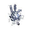
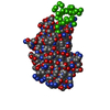
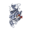
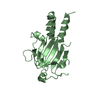
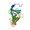
Journal: Open Biol / Year: 2014 Title: The basic keratin 10-binding domain of the virulence-associated pneumococcal serine-rich protein PsrP adopts a novel MSCRAMM fold. Authors: Tim Schulte / Jonas Löfling / Cecilia Mikaelsson / Alexey Kikhney / Karina Hentrich / Aurora Diamante / Christine Ebel / Staffan Normark / Dmitri Svergun / Birgitta Henriques-Normark / Adnane Achour / Abstract: Streptococcus pneumoniae is a major human pathogen, and a leading cause of disease and death worldwide. Pneumococcal invasive disease is triggered by initial asymptomatic colonization of the human ...Streptococcus pneumoniae is a major human pathogen, and a leading cause of disease and death worldwide. Pneumococcal invasive disease is triggered by initial asymptomatic colonization of the human upper respiratory tract. The pneumococcal serine-rich repeat protein (PsrP) is a lung-specific virulence factor whose functional binding region (BR) binds to keratin-10 (KRT10) and promotes pneumococcal biofilm formation through self-oligomerization. We present the crystal structure of the KRT10-binding domain of PsrP (BR187-385) determined to 2.0 Å resolution. BR187-385 adopts a novel variant of the DEv-IgG fold, typical for microbial surface components recognizing adhesive matrix molecules adhesins, despite very low sequence identity. An extended β-sheet on one side of the compressed, two-sided barrel presents a basic groove that possibly binds to the acidic helical rod domain of KRT10. Our study also demonstrates the importance of the other side of the barrel, formed by extensive well-ordered loops and stabilized by short β-strands, for interaction with KRT10.
History
Deposition
Dec 17, 2012
Deposition site: PDBE / Processing site: PDBE
Revision 1.0
Jan 8, 2014
Provider: repository / Type: Initial release
Revision 1.1
Jan 29, 2014
Group: Database references
Revision 1.2
May 8, 2019
Group: Advisory / Data collection ...Advisory / Data collection / Experimental preparation / Other Category: exptl_crystal_grow / pdbx_database_proc ...exptl_crystal_grow / pdbx_database_proc / pdbx_database_status / pdbx_unobs_or_zero_occ_atoms Item: _exptl_crystal_grow.method / _pdbx_database_status.recvd_author_approval
Mass: 18.015 Da / Num. of mol.: 114 / Source method: isolated from a natural source / Formula: H2O
-
Experimental details
-
Experiment
Experiment
Method: X-RAY DIFFRACTION / Number of used crystals: 1
-
Sample preparation
Crystal
Density Matthews: 1.9 Å3/Da / Density % sol: 35.35 % / Description: NONE
Crystal grow
Method: vapor diffusion, sitting drop Details: WELL-DIFFRACTING CRYSTALS OF WILD-TYPE AND SE-MET-BR187-385 WERE OBTAINED IN 0.2 M LITHIUM SULFATE, 0.1 M SODIUM ACETATE TRIHYDRATE PH 4.6, 25% PEG4000 (W/V) USING THE SITTING DROP VAPOR- ...Details: WELL-DIFFRACTING CRYSTALS OF WILD-TYPE AND SE-MET-BR187-385 WERE OBTAINED IN 0.2 M LITHIUM SULFATE, 0.1 M SODIUM ACETATE TRIHYDRATE PH 4.6, 25% PEG4000 (W/V) USING THE SITTING DROP VAPOR-DIFFUSION METHOD FOLLOWED BY MICRO-SEEDING.
Protocol: SINGLE WAVELENGTH / Monochromatic (M) / Laue (L): M / Scattering type: x-ray
Radiation wavelength
Wavelength: 0.977 Å / Relative weight: 1
Reflection
Resolution: 2→48.3 Å / Num. obs: 44053 / % possible obs: 100 % / Observed criterion σ(I): 3 / Redundancy: 6.7 % / Biso Wilson estimate: 35.34 Å2 / Rmerge(I) obs: 0.06 / Net I/σ(I): 18.2
Reflection shell
Resolution: 2→2.05 Å / Redundancy: 6.7 % / Rmerge(I) obs: 0.7 / Mean I/σ(I) obs: 3 / % possible all: 100
-
Processing
Software
Name
Version
Classification
PHENIX
(PHENIX.REFINE)
refinement
XDS
datareduction
XSCALE
datascaling
PHASER
phasing
Refinement
Method to determine structure: MOLECULAR REPLACEMENT Starting model: MODEL OBTAINED FROM SAD EXPERIMENT Resolution: 2→48.318 Å / SU ML: 0.61 / σ(F): 1.31 / Phase error: 16.43 / Stereochemistry target values: ML Details: A SINGLE RAMACHANDRAN PLOT OUTLIER WAS FOUND IN THE FINAL MODEL CORRESPONDING TO RESIDUE T271, LOCATED IN A LOOP REGION WITH WEAK ELECTRON DENSITY. RESIDUES T311, Q312 AND G313 ARE LOCALIZED ...Details: A SINGLE RAMACHANDRAN PLOT OUTLIER WAS FOUND IN THE FINAL MODEL CORRESPONDING TO RESIDUE T271, LOCATED IN A LOOP REGION WITH WEAK ELECTRON DENSITY. RESIDUES T311, Q312 AND G313 ARE LOCALIZED AT THE BEGINNING OF A TURN MOTIF WHICH WAS DIFFICULT TO MODEL. RESIDUES S376 AND S377 ARE LOCALIZED AT THE C-TERMINUS OF THE PROTEIN WITH WEAK ELECTRON DENSITY.
Rfactor
Num. reflection
% reflection
Rfree
0.201
2176
4.9 %
Rwork
0.1769
-
-
obs
0.1781
44052
99.95 %
Solvent computation
Shrinkage radii: 0.98 Å / VDW probe radii: 1.2 Å / Solvent model: FLAT BULK SOLVENT MODEL / Bsol: 51.984 Å2 / ksol: 0.373 e/Å3
Displacement parameters
Biso mean: 42.5 Å2
Baniso -1
Baniso -2
Baniso -3
1-
0 Å2
0 Å2
0 Å2
2-
-
0 Å2
0 Å2
3-
-
-
0 Å2
Refinement step
Cycle: LAST / Resolution: 2→48.318 Å
Protein
Nucleic acid
Ligand
Solvent
Total
Num. atoms
1352
0
28
114
1494
Refine LS restraints
Refine-ID
Type
Dev ideal
Number
X-RAY DIFFRACTION
f_bond_d
0.012
1403
X-RAY DIFFRACTION
f_angle_d
1.417
1896
X-RAY DIFFRACTION
f_dihedral_angle_d
13.242
494
X-RAY DIFFRACTION
f_chiral_restr
0.09
211
X-RAY DIFFRACTION
f_plane_restr
0.006
239
LS refinement shell
Resolution (Å)
Rfactor Rfree
Num. reflection Rfree
Rfactor Rwork
Num. reflection Rwork
Refine-ID
% reflection obs (%)
1.9999-2.0434
0.2896
133
0.2604
2593
X-RAY DIFFRACTION
100
2.0434-2.0909
0.2142
134
0.2408
2642
X-RAY DIFFRACTION
100
2.0909-2.1432
0.1921
139
0.2074
2611
X-RAY DIFFRACTION
100
2.1432-2.2012
0.2091
136
0.1832
2639
X-RAY DIFFRACTION
100
2.2012-2.2659
0.2004
131
0.1741
2619
X-RAY DIFFRACTION
100
2.2659-2.3391
0.1963
141
0.1675
2625
X-RAY DIFFRACTION
100
2.3391-2.4227
0.2224
138
0.1749
2581
X-RAY DIFFRACTION
100
2.4227-2.5197
0.1948
137
0.1673
2613
X-RAY DIFFRACTION
100
2.5197-2.6343
0.196
140
0.1777
2630
X-RAY DIFFRACTION
100
2.6343-2.7732
0.1871
136
0.1575
2621
X-RAY DIFFRACTION
100
2.7732-2.9469
0.1898
137
0.1649
2608
X-RAY DIFFRACTION
100
2.9469-3.1744
0.2026
142
0.1746
2608
X-RAY DIFFRACTION
100
3.1744-3.4938
0.189
135
0.1759
2628
X-RAY DIFFRACTION
100
3.4938-3.9992
0.1717
132
0.167
2605
X-RAY DIFFRACTION
100
3.9992-5.0377
0.1796
133
0.1442
2622
X-RAY DIFFRACTION
100
5.0377-48.332
0.2603
132
0.2174
2631
X-RAY DIFFRACTION
100
Refinement TLS params.
Method: refined / Origin x: -15.895 Å / Origin y: -34.4247 Å / Origin z: -4.5657 Å
11
12
13
21
22
23
31
32
33
T
0.1409 Å2
0.0471 Å2
-0.0278 Å2
-
0.2274 Å2
0.0212 Å2
-
-
0.2305 Å2
L
2.6323 °2
0.4247 °2
1.354 °2
-
1.7884 °2
1.0174 °2
-
-
4.1183 °2
S
0.105 Å °
-0.1908 Å °
-0.1624 Å °
0.0519 Å °
0.0793 Å °
-0.0784 Å °
0.3283 Å °
0.4245 Å °
-0.1281 Å °
Refinement TLS group
Selection details: (CHAIN A AND RESID 203:379)
+
About Yorodumi
-
News
-
Feb 9, 2022. New format data for meta-information of EMDB entries
New format data for meta-information of EMDB entries
Version 3 of the EMDB header file is now the official format.
The previous official version 1.9 will be removed from the archive.
In the structure databanks used in Yorodumi, some data are registered as the other names, "COVID-19 virus" and "2019-nCoV". Here are the details of the virus and the list of structure data.
Jan 31, 2019. EMDB accession codes are about to change! (news from PDBe EMDB page)
EMDB accession codes are about to change! (news from PDBe EMDB page)
The allocation of 4 digits for EMDB accession codes will soon come to an end. Whilst these codes will remain in use, new EMDB accession codes will include an additional digit and will expand incrementally as the available range of codes is exhausted. The current 4-digit format prefixed with “EMD-” (i.e. EMD-XXXX) will advance to a 5-digit format (i.e. EMD-XXXXX), and so on. It is currently estimated that the 4-digit codes will be depleted around Spring 2019, at which point the 5-digit format will come into force.
The EM Navigator/Yorodumi systems omit the EMD- prefix.
Related info.:Q: What is EMD? / ID/Accession-code notation in Yorodumi/EM Navigator
Yorodumi is a browser for structure data from EMDB, PDB, SASBDB, etc.
This page is also the successor to EM Navigator detail page, and also detail information page/front-end page for Omokage search.
The word "yorodu" (or yorozu) is an old Japanese word meaning "ten thousand". "mi" (miru) is to see.
Related info.:EMDB / PDB / SASBDB / Comparison of 3 databanks / Yorodumi Search / Aug 31, 2016. New EM Navigator & Yorodumi / Yorodumi Papers / Jmol/JSmol / Function and homology information / Changes in new EM Navigator and Yorodumi
 Movie
Movie Controller

 Yorodumi
 Open data
 Basic information
 Components
 Keywords
 Function and homology information
 STREPTOCOCCUS PNEUMONIAE (bacteria)
 X-RAY DIFFRACTION /
  Authors
 Citation
 Structure visualization
 Downloads & links
 Other downloads
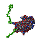


 PDBj
PDBj Assembly
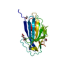

 Mass: 59.044 Da / Num. of mol.: 3 / Source method: obtained synthetically / Formula: C2H3O2
 Mass: 92.094 Da / Num. of mol.: 2 / Source method: obtained synthetically / Formula: C3H8O3
 Mass: 62.068 Da / Num. of mol.: 1 / Source method: obtained synthetically / Formula: C2H6O2
 Mass: 18.015 Da / Num. of mol.: 114 / Source method: isolated from a natural source / Formula: H2O
 Sample preparation
 / Beamline: ID29 / Wavelength: 0.977
  Processing