 Movie
Movie Controller


+ Open data

- Basic information
| Entry | Database: PDB / ID: 1jvw | ||||||
|---|---|---|---|---|---|---|---|
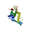
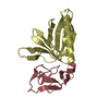
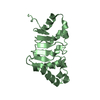
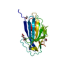
| Title | TRYPANOSOMA CRUZI MACROPHAGE INFECTIVITY POTENTIATOR (TCMIP) | ||||||
 Components Components | MACROPHAGE INFECTIVITY POTENTIATOR | ||||||
 Keywords Keywords | ISOMERASE / Macrophage Infectivity Potentiator / Trypanosoma cruzi / Chagas disease / X-ray crystal structure / rotamase | ||||||
| Function / homology |  Function and homology information Function and homology informationpeptidylprolyl isomerase / peptidyl-prolyl cis-trans isomerase activity / extracellular region Similarity search - Function | ||||||
| Biological species |  | ||||||
| Method |  X-RAY DIFFRACTION / SYNCHROTRON / MOLECULAR REPLACEMENT / Resolution: 1.7 Å X-RAY DIFFRACTION / SYNCHROTRON / MOLECULAR REPLACEMENT / Resolution: 1.7 Å | ||||||
 Authors Authors | Pereira, P.J.B. / Vega, M.C. / Gonzalez-Rey, E. / Fernandez-Carazo, R. / Macedo-Ribeiro, S. / Gomis-Rueth, F.X. / Gonzalez, A. / Coll, M. | ||||||
 Citation Citation | Journal: EMBO Rep. / Year: 2002 Title: Trypanosoma cruzi macrophage infectivity potentiator has a rotamase core and a highly exposed alpha-helix. Authors: Pereira, P.J. / Vega, M.C. / Gonzalez-Rey, E. / Fernandez-Carazo, R. / Macedo-Ribeiro, S. / Gomis-Ruth, F.X. / Gonzalez, A. / Coll, M. | ||||||
| History |
|
- Structure visualization
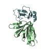
| Structure viewer | Molecule: MolmilJmol/JSmol |
|---|
- Downloads & links
-Download
| PDBx/mmCIF format | 1jvw.cif.gz | 50.4 KB | Display | PDBx/mmCIF format |
|---|---|---|---|---|
| PDB format | pdb1jvw.ent.gz | 35 KB | Display | PDB format |
| PDBx/mmJSON format | 1jvw.json.gz | Tree view | PDBx/mmJSON format | |
| Others |  Other downloads Other downloads |
-Validation report
| Arichive directory | https://data.pdbj.org/pub/pdb/validation_reports/jv/1jvwftp://data.pdbj.org/pub/pdb/validation_reports/jv/1jvw | HTTPS FTP |
|---|
-Related structure data
| Similar structure data |
|---|


-Links
 PDBj
PDBj

- Assembly
| Deposited unit | 
| ||||||||
|---|---|---|---|---|---|---|---|---|---|
| 1 |
| ||||||||
| Unit cell |
|
-Components
| #1: Protein | Mass: 18863.271 Da / Num. of mol.: 1 Source method: isolated from a genetically manipulated source Source: (gene. exp.)  |
|---|---|
| #2: Water | ChemComp-HOH /  Mass: 18.015 Da / Num. of mol.: 271 / Source method: isolated from a natural source / Formula: H2O Mass: 18.015 Da / Num. of mol.: 271 / Source method: isolated from a natural source / Formula: H2O |
-Experimental details
-Experiment
| Experiment | Method: X-RAY DIFFRACTION / Number of used crystals: 1 |
|---|
- Sample preparation
| Crystal | Density Matthews: 2.13 Å3/Da / Density % sol: 42.14 % | ||||||||||||||||||
|---|---|---|---|---|---|---|---|---|---|---|---|---|---|---|---|---|---|---|---|
| Crystal grow | Temperature: 277 K / Method: small tubes / pH: 7.5 Details: PMSF, Tris/HCl, pH 7.5, SMALL TUBES, temperature 277K | ||||||||||||||||||
| Crystal grow | *PLUS Temperature: 4 ℃ / Method: microdialysis | ||||||||||||||||||
| Components of the solutions | *PLUS
|
-Data collection
| Diffraction | Mean temperature: 100 K |
|---|---|
| Diffraction source | Source: SYNCHROTRON / Site: EMBL/DESY, HAMBURG  / Beamline: BW7B / Wavelength: 0.885 Å / Beamline: BW7B / Wavelength: 0.885 Å |
| Detector | Type: MARRESEARCH / Detector: IMAGE PLATE / Date: Dec 20, 1997 |
| Radiation | Monochromator: SAGITALLY FOCUSED Si(111) / Protocol: SINGLE WAVELENGTH / Monochromatic (M) / Laue (L): M / Scattering type: x-ray |
| Radiation wavelength | Wavelength: 0.885 Å / Relative weight: 1 |
| Reflection | Resolution: 1.7→19.2 Å / Num. obs: 17009 / % possible obs: 95.9 % / Observed criterion σ(F): 0 / Observed criterion σ(I): 0 |
| Reflection shell | Resolution: 1.7→1.79 Å / % possible all: 89.8 |
| Reflection | *PLUS Highest resolution: 1.7 Å / Redundancy: 3.6 % / Num. measured all: 60547 / Rmerge(I) obs: 0.081 |
| Reflection shell | *PLUS Highest resolution: 1.7 Å / % possible obs: 89.8 % / Rmerge(I) obs: 0.097 |
- Processing
| Software |
| ||||||||||||||||||||
|---|---|---|---|---|---|---|---|---|---|---|---|---|---|---|---|---|---|---|---|---|---|
| Refinement | Method to determine structure: MOLECULAR REPLACEMENT / Resolution: 1.7→19 Å / σ(F): 0 / Stereochemistry target values: Engh & Huber
| ||||||||||||||||||||
| Refinement step | Cycle: LAST / Resolution: 1.7→19 Å
| ||||||||||||||||||||
| Refine LS restraints |
| ||||||||||||||||||||
| Software | *PLUS Name: SHELXL / Version: 97 / Classification: refinement | ||||||||||||||||||||
| Refinement | *PLUS % reflection Rfree: 5 % / Rfactor all: 0.179 / Rfactor Rfree: 0.231 / Rfactor Rwork: 0.176 | ||||||||||||||||||||
| Solvent computation | *PLUS | ||||||||||||||||||||
| Displacement parameters | *PLUS |
