 Movie
Movie Controller
[English] 日本語
 Yorodumi
- SASDAR3: PsrP functional binding region (Functional binding region (187-38... -
+ Open data

- Basic information
| Entry | Database: SASBDB / ID: SASDAR3 |
|---|---|
 Sample Sample | PsrP functional binding region
|
| Function / homology |  Function and homology information Function and homology informationGram-positive-bacterium-type cell wall / single-species biofilm formation / symbiont-mediated perturbation of host defense response / cell adhesion / cell surface / DNA binding Similarity search - Function |
| Biological species |   Streptococcus pneumoniae (bacteria) Streptococcus pneumoniae (bacteria) |
 Citation Citation | Journal: Open Biol / Year: 2014 Title: The basic keratin 10-binding domain of the virulence-associated pneumococcal serine-rich protein PsrP adopts a novel MSCRAMM fold. Authors: Tim Schulte / Jonas Löfling / Cecilia Mikaelsson / Alexey Kikhney / Karina Hentrich / Aurora Diamante / Christine Ebel / Staffan Normark / Dmitri Svergun / Birgitta Henriques-Normark / Adnane Achour /  Abstract: Streptococcus pneumoniae is a major human pathogen, and a leading cause of disease and death worldwide. Pneumococcal invasive disease is triggered by initial asymptomatic colonization of the human ...Streptococcus pneumoniae is a major human pathogen, and a leading cause of disease and death worldwide. Pneumococcal invasive disease is triggered by initial asymptomatic colonization of the human upper respiratory tract. The pneumococcal serine-rich repeat protein (PsrP) is a lung-specific virulence factor whose functional binding region (BR) binds to keratin-10 (KRT10) and promotes pneumococcal biofilm formation through self-oligomerization. We present the crystal structure of the KRT10-binding domain of PsrP (BR187-385) determined to 2.0 Å resolution. BR187-385 adopts a novel variant of the DEv-IgG fold, typical for microbial surface components recognizing adhesive matrix molecules adhesins, despite very low sequence identity. An extended β-sheet on one side of the compressed, two-sided barrel presents a basic groove that possibly binds to the acidic helical rod domain of KRT10. Our study also demonstrates the importance of the other side of the barrel, formed by extensive well-ordered loops and stabilized by short β-strands, for interaction with KRT10. |
 Contact author Contact author |
|
- Structure visualization
| Structure viewer | Molecule: MolmilJmol/JSmol |
|---|
- Downloads & links
 SASDAR3

-Models
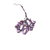
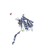
| Model #180 |  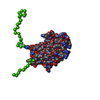 Type: mix / Software: EOM (1.3) / Radius of dummy atoms: 1.90 A / Chi-square value: 0.582169  Search similar-shape structures of this assembly by Omokage search (details) Search similar-shape structures of this assembly by Omokage search (details) |
|---|---|
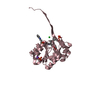
| Model #181 |  Type: mix / Software: EOM (1.3) / Radius of dummy atoms: 1.90 A / Chi-square value: 0.582169 Search similar-shape structures of this assembly by Omokage search (details) |
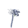
| Model #182 | 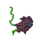 Type: mix / Software: EOM (1.3) / Radius of dummy atoms: 1.90 A / Chi-square value: 0.582169 Search similar-shape structures of this assembly by Omokage search (details) |
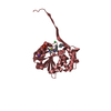
| Model #183 | 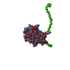 Type: mix / Software: EOM (1.3) / Radius of dummy atoms: 1.90 A / Chi-square value: 0.582169 Search similar-shape structures of this assembly by Omokage search (details) |
| Model #184 |  Type: mix / Software: EOM (1.3) / Radius of dummy atoms: 1.90 A / Chi-square value: 0.582169 Search similar-shape structures of this assembly by Omokage search (details) |
-Sample
| Sample | Name: PsrP functional binding region / Specimen concentration: 1.10-8.70 |
|---|---|
| Buffer | Name: sodium citrate / Concentration: 20.00 mM / pH: 5.5 / Comment: buffer for BR(187-385) / Composition: NaCl 250.000 mM, glycerol 2.500 % |
| Entity #37 | Name: PsrP BR(187-385) / Type: protein Description: Functional binding region (187-385) of the pneumococcal serine-rich repeat protein Formula weight: 22.12 / Num. of mol.: 1 / Source: Streptococcus pneumoniae / References: UniProt: A0A0H2URK1 Sequence: HHHHHHSGNT IVNGAPAINA SLNIAKSETK VYTGEGVDSV YRVPIYYKLK VTNDGSKLTF TYTVTYVNPK TNDLGNISSM RPGYSIYNSG TSTQTMLTLG SDLGKPSGVK NYITDKNGRQ VLSYNTSTMT TQGSGYTWGN GAQMNGFFAK KGYGLTSSWT VPITGTDTSF ...Sequence: HHHHHHSGNT IVNGAPAINA SLNIAKSETK VYTGEGVDSV YRVPIYYKLK VTNDGSKLTF TYTVTYVNPK TNDLGNISSM RPGYSIYNSG TSTQTMLTLG SDLGKPSGVK NYITDKNGRQ VLSYNTSTMT TQGSGYTWGN GAQMNGFFAK KGYGLTSSWT VPITGTDTSF TFTPYAARTD RIGINYFNGG GKVVESSTTS QSLSQ |
-Experimental information
| Beam | Instrument name:  DORIS III X33 DORIS III X33  / City: Hamburg / 国: Germany / Shape: 0.6 / Type of source: X-ray synchrotron / Wavelength: 0.15 Å / Dist. spec. to detc.: 2.7 mm / City: Hamburg / 国: Germany / Shape: 0.6 / Type of source: X-ray synchrotron / Wavelength: 0.15 Å / Dist. spec. to detc.: 2.7 mm | ||||||||||||||||||
|---|---|---|---|---|---|---|---|---|---|---|---|---|---|---|---|---|---|---|---|
| Detector | Name: Pilatus 1M-W / Pixsize x: 0.172 mm | ||||||||||||||||||
| Scan | 
| ||||||||||||||||||
| Distance distribution function P(R) | 
| ||||||||||||||||||
| Result | 
|
