 Movie
Movie Controller

[English] 日本語
 Yorodumi
- PDB-3zd1: STRUCTURE OF THE TWO C-TERMINAL DOMAINS OF COMPLEMENT FACTOR H RE... -
+ Open data

- Basic information
| Entry | Database: PDB / ID: 3zd1 | ||||||
|---|---|---|---|---|---|---|---|
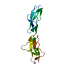
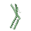
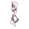
| Title | STRUCTURE OF THE TWO C-TERMINAL DOMAINS OF COMPLEMENT FACTOR H RELATED PROTEIN 2 | ||||||
 Components Components | COMPLEMENT FACTOR H-RELATED PROTEIN 2 | ||||||
 Keywords Keywords | IMMUNE SYSTEM / CFHR-2 / CFHR2 / FHR2 | ||||||
| Function / homology |  Function and homology information Function and homology informationcomplement component C3b binding / complement activation / : / negative regulation of protein binding / Regulation of Complement cascade / protein-containing complex / : / extracellular region / identical protein binding Similarity search - Function | ||||||
| Biological species |  HOMO SAPIENS (human) HOMO SAPIENS (human) | ||||||
| Method |  X-RAY DIFFRACTION / SYNCHROTRON / MOLECULAR REPLACEMENT / Resolution: 2 Å X-RAY DIFFRACTION / SYNCHROTRON / MOLECULAR REPLACEMENT / Resolution: 2 Å | ||||||
 Authors Authors | Caesar, J.J.E. / Goicoechea de Jorge, E. / Pickering, M.C. / Lea, S.M. | ||||||
 Citation Citation | Journal: Proc.Natl.Acad.Sci.USA / Year: 2013 Title: Dimerization of Complement Factor H-Related Proteins Modulates Complement Activation in Vivo. Authors: Goicoechea De Jorge, E. / Caesar, J.J.E. / Malik, T.H. / Patel, M. / Colledge, M. / Johnson, S. / Hakobyan, S. / Morgan, B.P. / Harris, C.L. / Pickering, M.C. / Lea, S.M. | ||||||
| History |
|
- Structure visualization
| Structure viewer | Molecule: MolmilJmol/JSmol |
|---|
- Downloads & links
-Download
| PDBx/mmCIF format | 3zd1.cif.gz | 64.1 KB | Display | PDBx/mmCIF format |
|---|---|---|---|---|
| PDB format | pdb3zd1.ent.gz | 47.2 KB | Display | PDB format |
| PDBx/mmJSON format | 3zd1.json.gz | Tree view | PDBx/mmJSON format | |
| Others |  Other downloads Other downloads |
-Validation report
| Arichive directory | https://data.pdbj.org/pub/pdb/validation_reports/zd/3zd1ftp://data.pdbj.org/pub/pdb/validation_reports/zd/3zd1 | HTTPS FTP |
|---|
-Related structure data
| Related structure data |  3zd2C 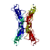 2g7iS C: citing same article ( S: Starting model for refinement |
|---|---|
| Similar structure data |


-Links
 PDBj
PDBj

- Assembly
| Deposited unit | 
| ||||||||
|---|---|---|---|---|---|---|---|---|---|
| 1 | 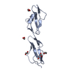
| ||||||||
| 2 | 
| ||||||||
| Unit cell |
| ||||||||
| Noncrystallographic symmetry (NCS) | NCS oper: (Code: given Matrix: (0.996773, -0.016503, -0.078563), Vector: |
-Components
| #1: Protein | Mass: 14303.232 Da / Num. of mol.: 2 / Fragment: SCR DOMAINS 3 AND 4, RESIDUES 147-270 Source method: isolated from a genetically manipulated source Source: (gene. exp.) HOMO SAPIENS (human) / Production host:  #2: Chemical | ChemComp-EDO /   Mass: 62.068 Da / Num. of mol.: 10 / Source method: obtained synthetically / Formula: C2H6O2 Mass: 62.068 Da / Num. of mol.: 10 / Source method: obtained synthetically / Formula: C2H6O2#3: Water | ChemComp-HOH / |  Mass: 18.015 Da / Num. of mol.: 77 / Source method: isolated from a natural source / Formula: H2O Mass: 18.015 Da / Num. of mol.: 77 / Source method: isolated from a natural source / Formula: H2OHas protein modification | Y | |
|---|
-Experimental details
-Experiment
| Experiment | Method: X-RAY DIFFRACTION / Number of used crystals: 1 |
|---|
- Sample preparation
| Crystal | Density Matthews: 2.23 Å3/Da / Density % sol: 44.96 % / Description: NONE |
|---|---|
| Crystal grow | Details: 30% (W/V) PEG 8000, 0.2M AMMONIUM SULPHATE |
-Data collection
| Diffraction | Mean temperature: 120 K |
|---|---|
| Diffraction source | Source: SYNCHROTRON / Site: Diamond  / Beamline: I04-1 / Wavelength: 0.9173 / Beamline: I04-1 / Wavelength: 0.9173 |
| Detector | Type: DECTRIS PILATUS 6M / Detector: PIXEL / Date: Dec 16, 2011 / Details: MIRRORS |
| Radiation | Monochromator: SINGLE BOUNCE / Protocol: SINGLE WAVELENGTH / Monochromatic (M) / Laue (L): M / Scattering type: x-ray |
| Radiation wavelength | Wavelength: 0.9173 Å / Relative weight: 1 |
| Reflection | Resolution: 2→95.47 Å / Num. obs: 16993 / % possible obs: 96.7 % / Observed criterion σ(I): 2 / Redundancy: 3.2 % / Biso Wilson estimate: 27.21 Å2 / Rmerge(I) obs: 0.05 / Net I/σ(I): 15.2 |
| Reflection shell | Resolution: 2→2.05 Å / Redundancy: 2.6 % / Rmerge(I) obs: 0.26 / Mean I/σ(I) obs: 4 / % possible all: 86.8 |
- Processing
| Software |
| ||||||||||||||||||||||||||||||||||||||||||||||||||||||||||||||||||||||||||||||||||||||||||||||||||||||||||||||||||
|---|---|---|---|---|---|---|---|---|---|---|---|---|---|---|---|---|---|---|---|---|---|---|---|---|---|---|---|---|---|---|---|---|---|---|---|---|---|---|---|---|---|---|---|---|---|---|---|---|---|---|---|---|---|---|---|---|---|---|---|---|---|---|---|---|---|---|---|---|---|---|---|---|---|---|---|---|---|---|---|---|---|---|---|---|---|---|---|---|---|---|---|---|---|---|---|---|---|---|---|---|---|---|---|---|---|---|---|---|---|---|---|---|---|---|---|
| Refinement | Method to determine structure: MOLECULAR REPLACEMENT Starting model: PDB ENTRY 2G7I Resolution: 2→19.09 Å / Cor.coef. Fo:Fc: 0.9283 / Cor.coef. Fo:Fc free: 0.9205 / SU R Cruickshank DPI: 0.207 / Cross valid method: THROUGHOUT / σ(F): 0 / SU R Blow DPI: 0.213 / SU Rfree Blow DPI: 0.17 / SU Rfree Cruickshank DPI: 0.169
| ||||||||||||||||||||||||||||||||||||||||||||||||||||||||||||||||||||||||||||||||||||||||||||||||||||||||||||||||||
| Displacement parameters | Biso mean: 27.45 Å2
| ||||||||||||||||||||||||||||||||||||||||||||||||||||||||||||||||||||||||||||||||||||||||||||||||||||||||||||||||||
| Refine analyze | Luzzati coordinate error obs: 0.241 Å | ||||||||||||||||||||||||||||||||||||||||||||||||||||||||||||||||||||||||||||||||||||||||||||||||||||||||||||||||||
| Refinement step | Cycle: LAST / Resolution: 2→19.09 Å
| ||||||||||||||||||||||||||||||||||||||||||||||||||||||||||||||||||||||||||||||||||||||||||||||||||||||||||||||||||
| Refine LS restraints |
| ||||||||||||||||||||||||||||||||||||||||||||||||||||||||||||||||||||||||||||||||||||||||||||||||||||||||||||||||||
| LS refinement shell | Resolution: 2→2.12 Å / Total num. of bins used: 9
|
