 Movie
Movie Controller


+ Open data

- Basic information
| Entry | Database: PDB / ID: 3kxv | ||||||
|---|---|---|---|---|---|---|---|
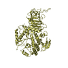
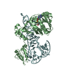
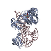
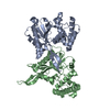
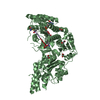
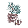
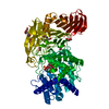
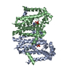
| Title | Structure of complement Factor H variant Q1139A | ||||||
 Components Components | Complement factor H | ||||||
 Keywords Keywords | IMMUNE SYSTEM / Sushi domain / short consensus repeat domain / SCR domain / complement control protein module / complement regulator / atypical hemolytic uremic syndrome / renal disease / CFH / complement alternative pathway / disease mutation / glycoprotein / immune response / innate immunity | ||||||
| Function / homology |  Function and homology information Function and homology informationregulation of complement activation, alternative pathway / regulation of complement-dependent cytotoxicity / regulation of complement activation / complement component C3b binding / central nervous system myelination / symbiont cell surface / serine-type endopeptidase complex / heparan sulfate proteoglycan binding / complement activation, alternative pathway / complement activation ...regulation of complement activation, alternative pathway / regulation of complement-dependent cytotoxicity / regulation of complement activation / complement component C3b binding / central nervous system myelination / symbiont cell surface / serine-type endopeptidase complex / heparan sulfate proteoglycan binding / complement activation, alternative pathway / complement activation / Regulation of Complement cascade / heparin binding / blood microparticle / inflammatory response / proteolysis / : / extracellular exosome / extracellular region / identical protein binding Similarity search - Function | ||||||
| Biological species |  Homo sapiens (human) Homo sapiens (human) | ||||||
| Method |  X-RAY DIFFRACTION / SYNCHROTRON / MOLECULAR REPLACEMENT / Resolution: 2.004 Å X-RAY DIFFRACTION / SYNCHROTRON / MOLECULAR REPLACEMENT / Resolution: 2.004 Å | ||||||
 Authors Authors | Bhattacharjee, A. / Lehtinen, M.J. / Kajander, T. / Goldman, A. / Jokiranta, T.S. | ||||||
 Citation Citation | Journal: Mol.Immunol. / Year: 2010 Title: Both domain 19 and domain 20 of factor H are involved in binding to complement C3b and C3d Authors: Bhattacharjee, A. / Lehtinen, M.J. / Kajander, T. / Goldman, A. / Jokiranta, T.S. | ||||||
| History |
|
- Structure visualization
| Structure viewer | Molecule: MolmilJmol/JSmol |
|---|
- Downloads & links
-Download
| PDBx/mmCIF format | 3kxv.cif.gz | 42.4 KB | Display | PDBx/mmCIF format |
|---|---|---|---|---|
| PDB format | pdb3kxv.ent.gz | 28.9 KB | Display | PDB format |
| PDBx/mmJSON format | 3kxv.json.gz | Tree view | PDBx/mmJSON format | |
| Others |  Other downloads Other downloads |
-Validation report
| Arichive directory | https://data.pdbj.org/pub/pdb/validation_reports/kx/3kxvftp://data.pdbj.org/pub/pdb/validation_reports/kx/3kxv | HTTPS FTP |
|---|
-Related structure data
| Related structure data |  3kzjC 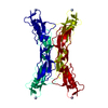 2g7iS S: Starting model for refinement C: citing same article ( |
|---|---|
| Similar structure data |


-Links
 PDBj
PDBj

- Assembly
| Deposited unit | 
| ||||||||
|---|---|---|---|---|---|---|---|---|---|
| 1 | 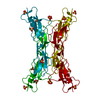
| ||||||||
| Unit cell |
|
-Components
| #1: Protein | Mass: 15042.128 Da / Num. of mol.: 1 / Fragment: Sushi domains 19-20 / Mutation: Q1139A Source method: isolated from a genetically manipulated source Source: (gene. exp.) Homo sapiens (human) / Plasmid: pPICZ-alpha- B / Production host:  Pichia pastoris (fungus) / References: UniProt: P08603 Pichia pastoris (fungus) / References: UniProt: P08603 | ||||
|---|---|---|---|---|---|
| #2: Chemical | ChemComp-SO4 /   Mass: 96.063 Da / Num. of mol.: 6 / Source method: obtained synthetically / Formula: SO4 Mass: 96.063 Da / Num. of mol.: 6 / Source method: obtained synthetically / Formula: SO4#3: Water | ChemComp-HOH / |  Mass: 18.015 Da / Num. of mol.: 113 / Source method: isolated from a natural source / Formula: H2O Mass: 18.015 Da / Num. of mol.: 113 / Source method: isolated from a natural source / Formula: H2OHas protein modification | Y | |
-Experimental details
-Experiment
| Experiment | Method: X-RAY DIFFRACTION / Number of used crystals: 1 |
|---|
- Sample preparation
| Crystal | Density Matthews: 4.07 Å3/Da / Density % sol: 69.81 % |
|---|---|
| Crystal grow | Temperature: 295.15 K / Method: vapor diffusion, sitting drop / pH: 5.5 Details: 2M ammonium sulphate, 0.1M Bis-Tris buffer, pH 5.5, VAPOR DIFFUSION, SITTING DROP, temperature 295.15K |
-Data collection
| Diffraction | Mean temperature: 100 K |
|---|---|
| Diffraction source | Source: SYNCHROTRON / Site: ESRF  / Beamline: ID14-3 / Wavelength: 0.931 Å / Beamline: ID14-3 / Wavelength: 0.931 Å |
| Detector | Type: ADSC QUANTUM 4 / Detector: CCD / Date: Dec 3, 2007 |
| Radiation | Monochromator: Diamond (111), Ge(220) / Protocol: SINGLE WAVELENGTH / Monochromatic (M) / Laue (L): M / Scattering type: x-ray |
| Radiation wavelength | Wavelength: 0.931 Å / Relative weight: 1 |
| Reflection | Resolution: 2→50 Å / Num. obs: 16014 / % possible obs: 99.7 % / Observed criterion σ(F): 0 / Observed criterion σ(I): -3 / Redundancy: 7.116 % / Biso Wilson estimate: 23.4 Å2 / Rsym value: 0.097 / Net I/σ(I): 16.8 |
| Reflection shell | Resolution: 2→2.13 Å / Redundancy: 7.16 % / Mean I/σ(I) obs: 3.43 / Num. unique all: 18145 / Rsym value: 0.552 / % possible all: 98.8 |
- Processing
| Software |
| |||||||||||||||||||||||||||||||||||
|---|---|---|---|---|---|---|---|---|---|---|---|---|---|---|---|---|---|---|---|---|---|---|---|---|---|---|---|---|---|---|---|---|---|---|---|---|
| Refinement | Method to determine structure: MOLECULAR REPLACEMENT Starting model: PDB ENTRY 2G7I Resolution: 2.004→42.012 Å / Occupancy max: 1 / Occupancy min: 0.46 / FOM work R set: 0.844 / SU ML: 1.86 Isotropic thermal model: Isotropic individual temperature factors Cross valid method: THROUGHOUT / σ(F): 2 / Phase error: 22.27 / Stereochemistry target values: ML / Details: The structure was refined also with REFMAC 5.1
| |||||||||||||||||||||||||||||||||||
| Solvent computation | Shrinkage radii: 0.9 Å / VDW probe radii: 1.11 Å / Solvent model: FLAT BULK SOLVENT MODEL / Bsol: 50.504 Å2 / ksol: 0.403 e/Å3 | |||||||||||||||||||||||||||||||||||
| Displacement parameters | Biso max: 82.04 Å2 / Biso mean: 25.159 Å2 / Biso min: 11.39 Å2
| |||||||||||||||||||||||||||||||||||
| Refinement step | Cycle: LAST / Resolution: 2.004→42.012 Å
| |||||||||||||||||||||||||||||||||||
| Refine LS restraints |
| |||||||||||||||||||||||||||||||||||
| LS refinement shell | Refine-ID: X-RAY DIFFRACTION / Total num. of bins used: 6 / % reflection obs: 100 %
|
