 Movie
Movie Controller

[English] 日本語
 Yorodumi
- PDB-2wda: The X-ray structure of the Streptomyces coelicolor A3 Chondroitin... -
+ Open data

- Basic information
| Entry | Database: PDB / ID: 2wda | |||||||||
|---|---|---|---|---|---|---|---|---|---|---|
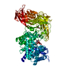
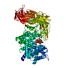
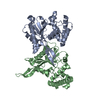
| Title | The X-ray structure of the Streptomyces coelicolor A3 Chondroitin AC Lyase in Complex with Chondroitin sulphate | |||||||||
 Components Components | PUTATIVE SECRETED LYASE | |||||||||
 Keywords Keywords | LYASE / HYALURONATE LYASE / CHONDROITIN LYASE / FAMILY 8 | |||||||||
| Function / homology |  Function and homology information Function and homology informationcarbon-oxygen lyase activity, acting on polysaccharides / carbohydrate binding / carbohydrate metabolic process / extracellular region / metal ion binding Similarity search - Function | |||||||||
| Biological species |  STREPTOMYCES VIOLACEORUBER (bacteria) STREPTOMYCES VIOLACEORUBER (bacteria) | |||||||||
| Method |  X-RAY DIFFRACTION / SYNCHROTRON / MOLECULAR REPLACEMENT / Resolution: 2.3 Å X-RAY DIFFRACTION / SYNCHROTRON / MOLECULAR REPLACEMENT / Resolution: 2.3 Å | |||||||||
 Authors Authors | Elmabrouk, Z.H. / Taylor, E.J. / Vincent, F. / Smith, N.L. / Turkenburg, J.P. / Davies, G.J. / Black, G.W. | |||||||||
 Citation Citation | Journal: Proteins / Year: 2011 Title: Crystal Structures of a Family 8 Polysaccharide Lyase Reveal Open and Highly Occluded Substrate-Binding Cleft Conformations. Authors: Elmabrouk, Z.H. / Vincent, F. / Zhang, M. / Smith, N.L. / Turkenburg, J.P. / Charnock, S.J. / Black, G.W. / Taylor, E.J. | |||||||||
| History |
|
- Structure visualization
| Structure viewer | Molecule: MolmilJmol/JSmol |
|---|
- Downloads & links
-Download
| PDBx/mmCIF format | 2wda.cif.gz | 170.8 KB | Display | PDBx/mmCIF format |
|---|---|---|---|---|
| PDB format | pdb2wda.ent.gz | 132.7 KB | Display | PDB format |
| PDBx/mmJSON format | 2wda.json.gz | Tree view | PDBx/mmJSON format | |
| Others |  Other downloads Other downloads |
-Validation report
| Arichive directory | https://data.pdbj.org/pub/pdb/validation_reports/wd/2wdaftp://data.pdbj.org/pub/pdb/validation_reports/wd/2wda | HTTPS FTP |
|---|
-Related structure data
| Related structure data |  2wcoSC  2x03C S: Starting model for refinement C: citing same article ( |
|---|---|
| Similar structure data |


-Links
 PDBj
PDBj


- Assembly
| Deposited unit | 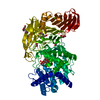
| ||||||||
|---|---|---|---|---|---|---|---|---|---|
| 1 |
| ||||||||
| Unit cell |
|
-Components
-Protein / Sugars , 2 types, 2 molecules A
| #1: Protein | Mass: 83022.234 Da / Num. of mol.: 1 / Fragment: RESIDUES 33-776 Source method: isolated from a genetically manipulated source Source: (gene. exp.) STREPTOMYCES VIOLACEORUBER (bacteria) / Strain: A3(2)Description: GERMAN COLLECTION OF MICROORGANISMS (DSM) DSM NO. 40783 Production host: |
|---|---|
| #2: Polysaccharide | 4-deoxy-alpha-L-threo-hex-4-enopyranuronic acid-(1-3)-2-acetamido-2-deoxy-4-O-sulfo-beta-D-galactopyranose Source method: isolated from a genetically manipulated source |
-Non-polymers , 5 types, 517 molecules 


| #3: Chemical | ChemComp-FMT /  Mass: 46.025 Da / Num. of mol.: 5 / Source method: obtained synthetically / Formula: CH2O2 Mass: 46.025 Da / Num. of mol.: 5 / Source method: obtained synthetically / Formula: CH2O2#4: Chemical |  Mass: 92.094 Da / Num. of mol.: 3 / Source method: obtained synthetically / Formula: C3H8O3 Mass: 92.094 Da / Num. of mol.: 3 / Source method: obtained synthetically / Formula: C3H8O3#5: Chemical | ChemComp-PEG /  Mass: 106.120 Da / Num. of mol.: 9 / Source method: obtained synthetically / Formula: C4H10O3 Mass: 106.120 Da / Num. of mol.: 9 / Source method: obtained synthetically / Formula: C4H10O3#6: Chemical | ChemComp-MG / |  Mass: 24.305 Da / Num. of mol.: 1 / Source method: obtained synthetically / Formula: Mg Mass: 24.305 Da / Num. of mol.: 1 / Source method: obtained synthetically / Formula: Mg#7: Water | ChemComp-HOH / | Mass: 18.015 Da / Num. of mol.: 499 / Source method: isolated from a natural source / Formula: H2O |
|---|
-Details
| Sequence details | THE CLONED SEQUENCE CONTAINS AN N_TERMINAL VECTOR DERIVED TAG FROM PET 28A, HOWEVER THIS IN NOT ...THE CLONED SEQUENCE CONTAINS AN N_TERMINAL VECTOR DERIVED TAG FROM PET 28A, HOWEVER THIS IN NOT ORDERED IN THE STRUCTURE. |
|---|
-Experimental details
-Experiment
| Experiment | Method: X-RAY DIFFRACTION / Number of used crystals: 1 |
|---|
- Sample preparation
| Crystal | Density Matthews: 3 Å3/Da / Density % sol: 59 % / Description: NONE |
|---|---|
| Crystal grow | Details: 4 M NA FORMATE,20 % GLYCEROL AS CRYOPROTECTANT |
-Data collection
| Diffraction | Mean temperature: 100 K |
|---|---|
| Diffraction source | Source: SYNCHROTRON / Site: ESRF  / Beamline: ID14-3 / Wavelength: 0.931 / Beamline: ID14-3 / Wavelength: 0.931 |
| Detector | Type: ADSC CCD / Detector: CCD / Date: May 7, 2005 |
| Radiation | Protocol: SINGLE WAVELENGTH / Monochromatic (M) / Laue (L): M / Scattering type: x-ray |
| Radiation wavelength | Wavelength: 0.931 Å / Relative weight: 1 |
| Reflection | Resolution: 2.3→50.3 Å / Num. obs: 45361 / % possible obs: 100 % / Observed criterion σ(I): 2 / Redundancy: 8.1 % / Rmerge(I) obs: 0.09 / Net I/σ(I): 7.6 |
| Reflection shell | Resolution: 2.3→2.4 Å / Redundancy: 8.2 % / Rmerge(I) obs: 0.39 / Mean I/σ(I) obs: 5 / % possible all: 100 |
- Processing
| Software |
| ||||||||||||||||||||||||||||||||||||||||||||||||||||||||||||||||||||||||||||||||||||||||||||||||||||||||||||||||||||||||||||||||||||||||||||||||||||||||||||||||||||||||||||||||||||||
|---|---|---|---|---|---|---|---|---|---|---|---|---|---|---|---|---|---|---|---|---|---|---|---|---|---|---|---|---|---|---|---|---|---|---|---|---|---|---|---|---|---|---|---|---|---|---|---|---|---|---|---|---|---|---|---|---|---|---|---|---|---|---|---|---|---|---|---|---|---|---|---|---|---|---|---|---|---|---|---|---|---|---|---|---|---|---|---|---|---|---|---|---|---|---|---|---|---|---|---|---|---|---|---|---|---|---|---|---|---|---|---|---|---|---|---|---|---|---|---|---|---|---|---|---|---|---|---|---|---|---|---|---|---|---|---|---|---|---|---|---|---|---|---|---|---|---|---|---|---|---|---|---|---|---|---|---|---|---|---|---|---|---|---|---|---|---|---|---|---|---|---|---|---|---|---|---|---|---|---|---|---|---|---|
| Refinement | Method to determine structure: MOLECULAR REPLACEMENT Starting model: PDB ENTRY 2WCO Resolution: 2.3→49.68 Å / Cor.coef. Fo:Fc: 0.962 / Cor.coef. Fo:Fc free: 0.942 / SU B: 4.27 / SU ML: 0.105 / Cross valid method: THROUGHOUT / ESU R: 0.209 / ESU R Free: 0.174 / Stereochemistry target values: MAXIMUM LIKELIHOOD Details: HYDROGENS HAVE BEEN ADDED IN THE RIDING POSITIONS. U VALUES REFINED INDIVIDUALLY
| ||||||||||||||||||||||||||||||||||||||||||||||||||||||||||||||||||||||||||||||||||||||||||||||||||||||||||||||||||||||||||||||||||||||||||||||||||||||||||||||||||||||||||||||||||||||
| Solvent computation | Ion probe radii: 0.8 Å / Shrinkage radii: 0.8 Å / VDW probe radii: 1.4 Å / Solvent model: MASK | ||||||||||||||||||||||||||||||||||||||||||||||||||||||||||||||||||||||||||||||||||||||||||||||||||||||||||||||||||||||||||||||||||||||||||||||||||||||||||||||||||||||||||||||||||||||
| Displacement parameters | Biso mean: 25.08 Å2
| ||||||||||||||||||||||||||||||||||||||||||||||||||||||||||||||||||||||||||||||||||||||||||||||||||||||||||||||||||||||||||||||||||||||||||||||||||||||||||||||||||||||||||||||||||||||
| Refinement step | Cycle: LAST / Resolution: 2.3→49.68 Å
| ||||||||||||||||||||||||||||||||||||||||||||||||||||||||||||||||||||||||||||||||||||||||||||||||||||||||||||||||||||||||||||||||||||||||||||||||||||||||||||||||||||||||||||||||||||||
| Refine LS restraints |
|