 Movie
Movie Controller

[English] 日本語
 Yorodumi
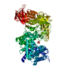
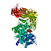
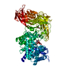
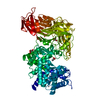
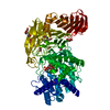
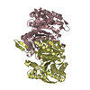
- PDB-1rwg: Crystal structure of Arthrobacter aurescens chondroitin AC lyase ... -
+ Open data

- Basic information
| Entry | Database: PDB / ID: 1rwg | |||||||||
|---|---|---|---|---|---|---|---|---|---|---|
| Title | Crystal structure of Arthrobacter aurescens chondroitin AC lyase in complex with chondroitin tetrasaccharide | |||||||||
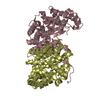
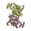
 Components Components | chondroitin AC lyase | |||||||||
 Keywords Keywords | LYASE / chondroitinase / chondroitin / chondroitin lyase | |||||||||
| Function / homology |  Function and homology information Function and homology informationcarbon-oxygen lyase activity, acting on polysaccharides / carbohydrate binding / carbohydrate metabolic process / extracellular region Similarity search - Function | |||||||||
| Biological species |  Arthrobacter aurescens (bacteria) Arthrobacter aurescens (bacteria) | |||||||||
| Method |  X-RAY DIFFRACTION / SYNCHROTRON / MOLECULAR REPLACEMENT / Resolution: 1.5 Å X-RAY DIFFRACTION / SYNCHROTRON / MOLECULAR REPLACEMENT / Resolution: 1.5 Å | |||||||||
 Authors Authors | Lunin, V.V. / Li, Y. / Miyazono, H. / Kyogashima, M. / Bell, A.W. / Cygler, M. | |||||||||
 Citation Citation | Journal: J.Mol.Biol. / Year: 2004 Title: High-resolution crystal structure of Arthrobacter aurescens chondroitin AC lyase: an enzyme-substrate complex defines the catalytic mechanism Authors: Lunin, V.V. / Li, Y. / Linhardt, R.J. / Miyazono, H. / Kyogashima, M. / Kaneko, T. / Bell, A.W. / Cygler, M. | |||||||||
| History |
| |||||||||
| Remark 600 | HETEROGEN SUGAR CHAIN D (GAD 809)-(NGL 810)-(GCT 811)-(NGL 812) IS CHONDROITIN SULFATE TETRASACCHARIDE. |
- Structure visualization
| Structure viewer | Molecule: MolmilJmol/JSmol |
|---|
- Downloads & links
-Download
| PDBx/mmCIF format | 1rwg.cif.gz | 336.2 KB | Display | PDBx/mmCIF format |
|---|---|---|---|---|
| PDB format | pdb1rwg.ent.gz | 269.9 KB | Display | PDB format |
| PDBx/mmJSON format | 1rwg.json.gz | Tree view | PDBx/mmJSON format | |
| Others |  Other downloads Other downloads |
-Validation report
| Arichive directory | https://data.pdbj.org/pub/pdb/validation_reports/rw/1rwgftp://data.pdbj.org/pub/pdb/validation_reports/rw/1rwg | HTTPS FTP |
|---|
-Related structure data
| Related structure data |  1rw9SC  1rwaC  1rwcC 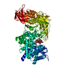 1rwfC  1rwhC S: Starting model for refinement C: citing same article ( |
|---|---|
| Similar structure data |


-Links
 PDBj
PDBj


- Assembly
| Deposited unit | 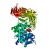
| ||||||||
|---|---|---|---|---|---|---|---|---|---|
| 1 |
| ||||||||
| Unit cell |
|
-Components
| #1: Protein | Mass: 79863.562 Da / Num. of mol.: 1 / Source method: isolated from a natural source / Source: (natural) Arthrobacter aurescens (bacteria) / References: UniProt: P84141, chondroitin AC lyase |
|---|---|
| #2: Polysaccharide | 2,6-anhydro-3-deoxy-L-threo-hex-2-enonic acid-(1-3)-2-acetamido-2-deoxy-4-O-sulfo-beta-D- ...2,6-anhydro-3-deoxy-L-threo-hex-2-enonic acid-(1-3)-2-acetamido-2-deoxy-4-O-sulfo-beta-D-galactopyranose-(1-4)-2,6-anhydro-3-deoxy-L-xylo-hexonic acid-(1-3)-2-acetamido-2-deoxy-4-O-sulfo-beta-D-galactopyranose Source method: isolated from a genetically manipulated source |
| #3: Chemical | ChemComp-NA /   Mass: 22.990 Da / Num. of mol.: 1 / Source method: obtained synthetically / Formula: Na Mass: 22.990 Da / Num. of mol.: 1 / Source method: obtained synthetically / Formula: Na |
| #4: Chemical | ChemComp-PO4 /   Mass: 94.971 Da / Num. of mol.: 1 / Source method: obtained synthetically / Formula: PO4 Mass: 94.971 Da / Num. of mol.: 1 / Source method: obtained synthetically / Formula: PO4 |
| #5: Water | ChemComp-HOH /  Mass: 18.015 Da / Num. of mol.: 1061 / Source method: isolated from a natural source / Formula: H2O Mass: 18.015 Da / Num. of mol.: 1061 / Source method: isolated from a natural source / Formula: H2O |
-Experimental details
-Experiment
| Experiment | Method: X-RAY DIFFRACTION / Number of used crystals: 1 |
|---|
- Sample preparation
| Crystal | Density Matthews: 2.41 Å3/Da / Density % sol: 48.88 % |
|---|---|
| Crystal grow | Method: vapor diffusion, hanging drop / pH: 6.4 Details: PEG 8000, ammonium acetate, glycerol, phosphate buffer, 10 minutes soaking time, pH 6.4, VAPOR DIFFUSION, HANGING DROP, temperature 100K |
-Data collection
| Diffraction | Mean temperature: 100 K |
|---|---|
| Diffraction source | Source: SYNCHROTRON / Site: NSLS  / Beamline: X8C / Wavelength: 0.9787 Å / Beamline: X8C / Wavelength: 0.9787 Å |
| Detector | Type: ADSC QUANTUM 4 / Detector: CCD / Date: Jun 28, 2002 |
| Radiation | Protocol: SINGLE WAVELENGTH / Monochromatic (M) / Laue (L): M / Scattering type: x-ray |
| Radiation wavelength | Wavelength: 0.9787 Å / Relative weight: 1 |
| Reflection | Resolution: 1.5→50 Å / Num. obs: 120769 / % possible obs: 99.9 % / Observed criterion σ(F): 1 / Observed criterion σ(I): 1 / Redundancy: 3.8 % / Rsym value: 0.077 / Net I/σ(I): 9.2 |
| Reflection shell | Resolution: 1.5→1.55 Å / % possible all: 99.9 |
- Processing
| Software |
| ||||||||||||||||||||||||||||||||||||||||||||||||||||||||||||||||||||||||||||||||||||||||||||||||||||||||||||||||||||||||||||||||||||||||||||||||||||||||||||||||
|---|---|---|---|---|---|---|---|---|---|---|---|---|---|---|---|---|---|---|---|---|---|---|---|---|---|---|---|---|---|---|---|---|---|---|---|---|---|---|---|---|---|---|---|---|---|---|---|---|---|---|---|---|---|---|---|---|---|---|---|---|---|---|---|---|---|---|---|---|---|---|---|---|---|---|---|---|---|---|---|---|---|---|---|---|---|---|---|---|---|---|---|---|---|---|---|---|---|---|---|---|---|---|---|---|---|---|---|---|---|---|---|---|---|---|---|---|---|---|---|---|---|---|---|---|---|---|---|---|---|---|---|---|---|---|---|---|---|---|---|---|---|---|---|---|---|---|---|---|---|---|---|---|---|---|---|---|---|---|---|---|---|
| Refinement | Method to determine structure: MOLECULAR REPLACEMENT Starting model: PDB entry 1RW9 Resolution: 1.5→28.06 Å / Cor.coef. Fo:Fc: 0.977 / Cor.coef. Fo:Fc free: 0.965 / SU B: 0.993 / SU ML: 0.037 / Cross valid method: THROUGHOUT / σ(F): 0 / ESU R: 0.07 / ESU R Free: 0.062 / Stereochemistry target values: MAXIMUM LIKELIHOOD
| ||||||||||||||||||||||||||||||||||||||||||||||||||||||||||||||||||||||||||||||||||||||||||||||||||||||||||||||||||||||||||||||||||||||||||||||||||||||||||||||||
| Solvent computation | Ion probe radii: 0.8 Å / Shrinkage radii: 0.8 Å / VDW probe radii: 1.4 Å / Solvent model: BABINET MODEL WITH MASK | ||||||||||||||||||||||||||||||||||||||||||||||||||||||||||||||||||||||||||||||||||||||||||||||||||||||||||||||||||||||||||||||||||||||||||||||||||||||||||||||||
| Displacement parameters | Biso mean: 18.336 Å2
| ||||||||||||||||||||||||||||||||||||||||||||||||||||||||||||||||||||||||||||||||||||||||||||||||||||||||||||||||||||||||||||||||||||||||||||||||||||||||||||||||
| Refinement step | Cycle: LAST / Resolution: 1.5→28.06 Å
| ||||||||||||||||||||||||||||||||||||||||||||||||||||||||||||||||||||||||||||||||||||||||||||||||||||||||||||||||||||||||||||||||||||||||||||||||||||||||||||||||
| Refine LS restraints |
| ||||||||||||||||||||||||||||||||||||||||||||||||||||||||||||||||||||||||||||||||||||||||||||||||||||||||||||||||||||||||||||||||||||||||||||||||||||||||||||||||
| LS refinement shell | Resolution: 1.5→1.539 Å / Total num. of bins used: 20 /
|
