Mass: 18.015 Da / Num. of mol.: 177 / Source method: isolated from a natural source / Formula: H2O
Has protein modification
Y
-
Experimental details
-
Experiment
Experiment
Method: X-RAY DIFFRACTION / Number of used crystals: 2
-
Sample preparation
Crystal
Density Matthews: 4.05 Å3/Da / Density % sol: 69.61 %
Crystal grow
Temperature: 298 K / Method: vapor diffusion, sitting drop / pH: 8.5 Details: Before crystallization the protein was concentrated to 10 mg/ml and dialysed into 20 mM Tris, 50 mM NaCl, pH 7.0. The protein was crystallized in sitting drops by mixing 1 ul of protein ...Details: Before crystallization the protein was concentrated to 10 mg/ml and dialysed into 20 mM Tris, 50 mM NaCl, pH 7.0. The protein was crystallized in sitting drops by mixing 1 ul of protein solution at 10 mg/ml with 1 ul of reservoir solution of 2.2 M (NH4)2SO4, 0.1 M Tris-HCl, pH 8.5, VAPOR DIFFUSION, SITTING DROP, temperature 298.0K
-
Data collection
Diffraction
ID
Mean temperature (K)
Crystal-ID
1
100
1
2
100
1
Diffraction source
Source
Site
Beamline
Type
ID
Wavelength (Å)
SYNCHROTRON
ESRF
ID14-1
1
0.934
ROTATING ANODE
RIGAKU RU300
2
1.54
Detector
Type
ID
Detector
Date
MARRESEARCH
1
CCD
Oct 14, 2003
RIGAKU RAXIS IV
2
IMAGE PLATE
Oct 20, 2003
Radiation
Protocol: SINGLE WAVELENGTH / Monochromatic (M) / Laue (L): M / Scattering type: x-ray
Resolution: 1.75→1.795 Å / Redundancy: 5.1 % / Mean I/σ(I) obs: 3.7 / Rsym value: 0.441 / % possible all: 99.9
-
Processing
Software
Name
Version
Classification
REFMAC
5.2.0005
refinement
XDS
datareduction
XDS
datascaling
SHELXS
phasing
Refinement
Method to determine structure: SAD / Resolution: 1.75→14.78 Å / Cor.coef. Fo:Fc: 0.942 / Cor.coef. Fo:Fc free: 0.935 / SU B: 1.631 / SU ML: 0.055 / Cross valid method: THROUGHOUT / σ(F): 0 / ESU R: 0.093 / ESU R Free: 0.09 / Stereochemistry target values: MAXIMUM LIKELIHOOD / Details: HYDROGENS HAVE BEEN ADDED IN THE RIDING POSITIONS
Rfactor
Num. reflection
% reflection
Selection details
Rfree
0.22479
1239
5.2 %
RANDOM
Rwork
0.20641
-
-
-
all
0.20735
22700
-
-
obs
0.20735
22700
99.93 %
-
Solvent computation
Ion probe radii: 0.8 Å / Shrinkage radii: 0.8 Å / VDW probe radii: 1.2 Å / Solvent model: MASK
Displacement parameters
Biso mean: 23.593 Å2
Baniso -1
Baniso -2
Baniso -3
1-
-0.06 Å2
0 Å2
0 Å2
2-
-
-0.06 Å2
0 Å2
3-
-
-
0.11 Å2
Refinement step
Cycle: LAST / Resolution: 1.75→14.78 Å
Protein
Nucleic acid
Ligand
Solvent
Total
Num. atoms
976
0
0
177
1153
Refine LS restraints
Refine-ID
Type
Dev ideal
Dev ideal target
Number
X-RAY DIFFRACTION
r_bond_refined_d
0.019
0.022
1001
X-RAY DIFFRACTION
r_bond_other_d
0.001
0.02
872
X-RAY DIFFRACTION
r_angle_refined_deg
1.645
1.963
1358
X-RAY DIFFRACTION
r_angle_other_deg
0.895
3
2038
X-RAY DIFFRACTION
r_dihedral_angle_1_deg
6.6
5
123
X-RAY DIFFRACTION
r_dihedral_angle_2_deg
33.646
24.146
41
X-RAY DIFFRACTION
r_dihedral_angle_3_deg
11.179
15
163
X-RAY DIFFRACTION
r_dihedral_angle_4_deg
10.057
15
5
X-RAY DIFFRACTION
r_chiral_restr
0.118
0.2
143
X-RAY DIFFRACTION
r_gen_planes_refined
0.009
0.02
1108
X-RAY DIFFRACTION
r_gen_planes_other
0.003
0.02
200
X-RAY DIFFRACTION
r_nbd_refined
0.213
0.2
202
X-RAY DIFFRACTION
r_nbd_other
0.181
0.2
827
X-RAY DIFFRACTION
r_nbtor_refined
0.176
0.2
477
X-RAY DIFFRACTION
r_nbtor_other
0.085
0.2
562
X-RAY DIFFRACTION
r_xyhbond_nbd_refined
0.161
0.2
101
X-RAY DIFFRACTION
r_symmetry_vdw_refined
0.254
0.2
17
X-RAY DIFFRACTION
r_symmetry_vdw_other
0.322
0.2
51
X-RAY DIFFRACTION
r_symmetry_hbond_refined
0.32
0.2
29
X-RAY DIFFRACTION
r_mcbond_it
1.453
1.5
671
X-RAY DIFFRACTION
r_mcbond_other
0.319
1.5
248
X-RAY DIFFRACTION
r_mcangle_it
2.08
2
1006
X-RAY DIFFRACTION
r_scbond_it
3.508
3
435
X-RAY DIFFRACTION
r_scangle_it
4.865
4.5
352
LS refinement shell
Resolution: 1.75→1.795 Å / Total num. of bins used: 20
Rfactor
Num. reflection
% reflection
Rfree
0.295
87
-
Rwork
0.279
1642
-
obs
-
-
100 %
+
About Yorodumi
-
News
-
Feb 9, 2022. New format data for meta-information of EMDB entries
New format data for meta-information of EMDB entries
Version 3 of the EMDB header file is now the official format.
The previous official version 1.9 will be removed from the archive.
In the structure databanks used in Yorodumi, some data are registered as the other names, "COVID-19 virus" and "2019-nCoV". Here are the details of the virus and the list of structure data.
Jan 31, 2019. EMDB accession codes are about to change! (news from PDBe EMDB page)
EMDB accession codes are about to change! (news from PDBe EMDB page)
The allocation of 4 digits for EMDB accession codes will soon come to an end. Whilst these codes will remain in use, new EMDB accession codes will include an additional digit and will expand incrementally as the available range of codes is exhausted. The current 4-digit format prefixed with “EMD-” (i.e. EMD-XXXX) will advance to a 5-digit format (i.e. EMD-XXXXX), and so on. It is currently estimated that the 4-digit codes will be depleted around Spring 2019, at which point the 5-digit format will come into force.
The EM Navigator/Yorodumi systems omit the EMD- prefix.
Related info.:Q: What is EMD? / ID/Accession-code notation in Yorodumi/EM Navigator
Yorodumi is a browser for structure data from EMDB, PDB, SASBDB, etc.
This page is also the successor to EM Navigator detail page, and also detail information page/front-end page for Omokage search.
The word "yorodu" (or yorozu) is an old Japanese word meaning "ten thousand". "mi" (miru) is to see.
Related info.:EMDB / PDB / SASBDB / Comparison of 3 databanks / Yorodumi Search / Aug 31, 2016. New EM Navigator & Yorodumi / Yorodumi Papers / Jmol/JSmol / Function and homology information / Changes in new EM Navigator and Yorodumi
 Movie
Movie Controller

 Yorodumi
 Open data
 Basic information
 Components
 Keywords
 Function and homology information
 Homo sapiens (human)
 X-RAY DIFFRACTION /
  Authors
 Citation
 Structure visualization
 Downloads & links
 Other downloads
 PDBj
PDBj

 Assembly
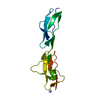
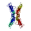
 Pichia pastoris (fungus) / References: UniProt: P08603
 Mass: 18.015 Da / Num. of mol.: 177 / Source method: isolated from a natural source / Formula: H2O
 Sample preparation
 Processing