 Movie
Movie Controller

[English] 日本語
 Yorodumi
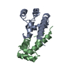
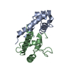
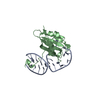
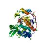
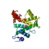
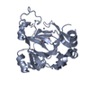
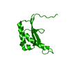
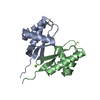
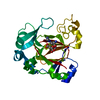
- PDB-3x0g: Crystal structure of the ectodomain of African green monkey CD81 ... -
+ Open data

- Basic information
| Entry | Database: PDB / ID: 3x0g | ||||||
|---|---|---|---|---|---|---|---|
| Title | Crystal structure of the ectodomain of African green monkey CD81 large extracellular loop (agmCD81-LEL) | ||||||
 Components Components | CD81 | ||||||
 Keywords Keywords | CELL ADHESION / disulfide bond / helical bundle / immune cell adhesion / morphology / activation / proliferation / differentiation / Membrane | ||||||
| Function / homology |  Function and homology information Function and homology informationpositive regulation of adaptive immune memory response / positive regulation of protein catabolic process in the vacuole / CD4-positive, alpha-beta T cell costimulation / positive regulation of B cell receptor signaling pathway / osteoclast fusion / myoblast fusion involved in skeletal muscle regeneration / positive regulation of T cell activation via T cell receptor contact with antigen bound to MHC molecule on antigen presenting cell / positive regulation of inflammatory response to antigenic stimulus / regulation of macrophage migration / macrophage fusion ...positive regulation of adaptive immune memory response / positive regulation of protein catabolic process in the vacuole / CD4-positive, alpha-beta T cell costimulation / positive regulation of B cell receptor signaling pathway / osteoclast fusion / myoblast fusion involved in skeletal muscle regeneration / positive regulation of T cell activation via T cell receptor contact with antigen bound to MHC molecule on antigen presenting cell / positive regulation of inflammatory response to antigenic stimulus / regulation of macrophage migration / macrophage fusion / immunological synapse formation / tetraspanin-enriched microdomain / positive regulation of T-helper 2 cell cytokine production / transferrin receptor binding / humoral immune response mediated by circulating immunoglobulin / positive regulation of protein exit from endoplasmic reticulum / protein localization to lysosome / MHC class II protein binding / positive regulation of CD4-positive, alpha-beta T cell proliferation / positive regulation of T cell receptor signaling pathway / cholesterol binding / immunological synapse / cellular response to low-density lipoprotein particle stimulus / positive regulation of receptor clustering / positive regulation of B cell proliferation / protein localization to plasma membrane / regulation of protein stability / receptor internalization / integrin binding / virus receptor activity / basolateral plasma membrane / positive regulation of MAPK cascade / positive regulation of transcription by RNA polymerase II / extracellular exosome / cytosol Similarity search - Function | ||||||
| Biological species |  Chlorocebus sabaeus (green monkey) Chlorocebus sabaeus (green monkey) | ||||||
| Method |  X-RAY DIFFRACTION / MOLECULAR REPLACEMENT / Resolution: 1.899 Å X-RAY DIFFRACTION / MOLECULAR REPLACEMENT / Resolution: 1.899 Å | ||||||
 Authors Authors | Zhang, M. / Cui, S. | ||||||
 Citation Citation | Journal: Faseb J. / Year: 2015 Title: An intramolecular bond at cluster of differentiation 81 ectodomain is important for hepatitis C virus entry. Authors: Yang, W. / Zhang, M. / Chi, X. / Liu, X. / Qin, B. / Cui, S. | ||||||
| History |
|
- Structure visualization
| Structure viewer | Molecule: MolmilJmol/JSmol |
|---|
- Downloads & links
-Download
| PDBx/mmCIF format | 3x0g.cif.gz | 46.9 KB | Display | PDBx/mmCIF format |
|---|---|---|---|---|
| PDB format | pdb3x0g.ent.gz | 34.4 KB | Display | PDB format |
| PDBx/mmJSON format | 3x0g.json.gz | Tree view | PDBx/mmJSON format | |
| Others |  Other downloads Other downloads |
-Validation report
| Arichive directory | https://data.pdbj.org/pub/pdb/validation_reports/x0/3x0gftp://data.pdbj.org/pub/pdb/validation_reports/x0/3x0g | HTTPS FTP |
|---|
-Related structure data
| Related structure data | 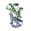 3x0eC  3x0fC  4bkh C: citing same article ( S: Starting model for refinement |
|---|---|
| Similar structure data |

-Links
 PDBj
PDBj- Assembly
| Deposited unit | 
| ||||||||
|---|---|---|---|---|---|---|---|---|---|
| 1 | 
| ||||||||
| Unit cell |
|
-Components
| #1: Protein | Mass: 10204.521 Da / Num. of mol.: 1 / Fragment: large extracellular loop Source method: isolated from a genetically manipulated source Source: (gene. exp.) Chlorocebus sabaeus (green monkey) / Gene: CD81 / Plasmid: pET-28a / Production host:  | ||||||
|---|---|---|---|---|---|---|---|
| #2: Chemical |   Mass: 118.174 Da / Num. of mol.: 3 / Source method: obtained synthetically / Formula: C6H14O2 / Comment: precipitant*YM Mass: 118.174 Da / Num. of mol.: 3 / Source method: obtained synthetically / Formula: C6H14O2 / Comment: precipitant*YM#3: Water | ChemComp-HOH / |  Mass: 18.015 Da / Num. of mol.: 56 / Source method: isolated from a natural source / Formula: H2O Mass: 18.015 Da / Num. of mol.: 56 / Source method: isolated from a natural source / Formula: H2OHas protein modification | Y | Sequence details | THE SEQUENCE OF THIS PROTEIN WAS NOT AVAILABLE AT THE UNIPROT KNOWLEDGEBASE DATABASE (UNIPROTKB) AT ...THE SEQUENCE OF THIS PROTEIN WAS NOT AVAILABLE AT THE UNIPROT KNOWLEDGEB | |
-Experimental details
-Experiment
| Experiment | Method: X-RAY DIFFRACTION / Number of used crystals: 1 |
|---|
- Sample preparation
| Crystal | Density Matthews: 2.28 Å3/Da / Density % sol: 46.09 % |
|---|---|
| Crystal grow | Temperature: 295 K / Method: vapor diffusion, hanging drop / pH: 8.5 Details: 5% Ethanol, 5% MPD, 0.1M Tris pH 8.5, 200mM Sodium Chloride, VAPOR DIFFUSION, HANGING DROP, temperature 295K |
-Data collection
| Diffraction | Mean temperature: 100 K |
|---|---|
| Diffraction source | Source: ROTATING ANODE / Type: RIGAKU MICROMAX-007 HF / Wavelength: 1.54 Å |
| Detector | Type: RIGAKU SATURN 944+ / Detector: CCD / Date: Jun 27, 2014 / Details: mirrors |
| Radiation | Monochromator: double crystal Si(111) / Protocol: SINGLE WAVELENGTH / Monochromatic (M) / Laue (L): M / Scattering type: x-ray |
| Radiation wavelength | Wavelength: 1.54 Å / Relative weight: 1 |
| Reflection | Resolution: 1.899→27.878 Å / Num. all: 7800 / Num. obs: 7580 / % possible obs: 97.2 % / Observed criterion σ(F): -3 / Observed criterion σ(I): -3 / Redundancy: 5.93 % / Biso Wilson estimate: 31.196 Å2 / Rmerge(I) obs: 0.064 / Net I/σ(I): 21.28 |
| Reflection shell | Resolution: 1.9→2.01 Å / Redundancy: 2.52 % / Rmerge(I) obs: 0.485 / Mean I/σ(I) obs: 2.42 / Num. unique all: 1219 / % possible all: 83.1 |
- Processing
| Software |
| ||||||||||||||||||||||||
|---|---|---|---|---|---|---|---|---|---|---|---|---|---|---|---|---|---|---|---|---|---|---|---|---|---|
| Refinement | Method to determine structure: MOLECULAR REPLACEMENT Starting model: 4BKH 4bkh Resolution: 1.899→25.276 Å / SU ML: 0.19 / Cross valid method: THROUGHOUT / σ(F): 1.35 / Phase error: 28.44 / Stereochemistry target values: ML
| ||||||||||||||||||||||||
| Solvent computation | Shrinkage radii: 0.86 Å / VDW probe radii: 1.1 Å / Solvent model: FLAT BULK SOLVENT MODEL / Bsol: 33.585 Å2 / ksol: 0.327 e/Å3 | ||||||||||||||||||||||||
| Displacement parameters | Biso mean: 30.81 Å2
| ||||||||||||||||||||||||
| Refinement step | Cycle: LAST / Resolution: 1.899→25.276 Å
| ||||||||||||||||||||||||
| Refine LS restraints |
| ||||||||||||||||||||||||
| LS refinement shell | Refine-ID: X-RAY DIFFRACTION / Total num. of bins used: 2
|
