 Movie
Movie Controller

[English] 日本語
 Yorodumi
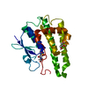
- PDB-6ax3: Complex structure of JMJD5 and Symmetric Dimethyl-Arginine (SDMA) -
+ Open data

- Basic information
| Entry | Database: PDB / ID: 6ax3 | ||||||
|---|---|---|---|---|---|---|---|
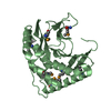
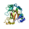
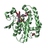
| Title | Complex structure of JMJD5 and Symmetric Dimethyl-Arginine (SDMA) | ||||||
 Components Components | Lysine-specific demethylase 8 | ||||||
 Keywords Keywords | HYDROLASE / Demethylase / Jumonji / Histone / Endopeptidase / Exopeptidase | ||||||
| Function / homology |  Function and homology information Function and homology information[protein]-arginine 3-hydroxylase / peptidyl-arginine 3-dioxygenase activity / histone H3K36 demethylase activity / Hydrolases; Acting on peptide bonds (peptidases) / Protein hydroxylation / aminopeptidase activity / regulation of signal transduction by p53 class mediator / circadian regulation of gene expression / protein destabilization / G2/M transition of mitotic cell cycle ...[protein]-arginine 3-hydroxylase / peptidyl-arginine 3-dioxygenase activity / histone H3K36 demethylase activity / Hydrolases; Acting on peptide bonds (peptidases) / Protein hydroxylation / aminopeptidase activity / regulation of signal transduction by p53 class mediator / circadian regulation of gene expression / protein destabilization / G2/M transition of mitotic cell cycle / p53 binding / chromosome / fibroblast proliferation / endopeptidase activity / in utero embryonic development / negative regulation of DNA-templated transcription / chromatin binding / positive regulation of DNA-templated transcription / proteolysis / nucleoplasm / metal ion binding / nucleus / cytosol Similarity search - Function | ||||||
| Biological species |  Homo sapiens (human) Homo sapiens (human) | ||||||
| Method |  X-RAY DIFFRACTION / SYNCHROTRON / MOLECULAR REPLACEMENT / Resolution: 2.25 Å X-RAY DIFFRACTION / SYNCHROTRON / MOLECULAR REPLACEMENT / Resolution: 2.25 Å | ||||||
 Authors Authors | Lee, S. / Liu, H. / Wang, Y. / Dai, S. / Zhang, G. | ||||||
 Citation Citation | Journal: Sci Rep / Year: 2018 Title: Specific Recognition of Arginine Methylated Histone Tails by JMJD5 and JMJD7. Authors: Liu, H. / Wang, C. / Lee, S. / Ning, F. / Wang, Y. / Zhang, Q. / Chen, Z. / Zang, J. / Nix, J. / Dai, S. / Marrack, P. / Hagman, J. / Kappler, J. / Zhang, G. | ||||||
| History |
|
- Structure visualization
| Structure viewer | Molecule: MolmilJmol/JSmol |
|---|
- Downloads & links
-Download
| PDBx/mmCIF format | 6ax3.cif.gz | 66.7 KB | Display | PDBx/mmCIF format |
|---|---|---|---|---|
| PDB format | pdb6ax3.ent.gz | 46.7 KB | Display | PDB format |
| PDBx/mmJSON format | 6ax3.json.gz | Tree view | PDBx/mmJSON format | |
| Others |  Other downloads Other downloads |
-Validation report
| Arichive directory | https://data.pdbj.org/pub/pdb/validation_reports/ax/6ax3ftp://data.pdbj.org/pub/pdb/validation_reports/ax/6ax3 | HTTPS FTP |
|---|
-Related structure data
| Related structure data |  6avsC 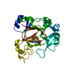 5fbjS S: Starting model for refinement C: citing same article ( |
|---|---|
| Similar structure data |


-Links
 PDBj
PDBj


- Assembly
| Deposited unit | 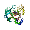
| ||||||||
|---|---|---|---|---|---|---|---|---|---|
| 1 |
| ||||||||
| Unit cell |
|
-Components
| #1: Protein | Mass: 27274.873 Da / Num. of mol.: 1 Source method: isolated from a genetically manipulated source Source: (gene. exp.) Homo sapiens (human) / Gene: KDM8, JMJD5Production host:  References: UniProt: Q8N371, [histone H3]-dimethyl-L-lysine36 demethylase |
|---|---|
| #2: Chemical | ChemComp-2MR /   Type: L-peptide linking / Mass: 202.254 Da / Num. of mol.: 1 / Source method: obtained synthetically / Formula: C8H18N4O2 Type: L-peptide linking / Mass: 202.254 Da / Num. of mol.: 1 / Source method: obtained synthetically / Formula: C8H18N4O2 |
| #3: Chemical | ChemComp-AKG /   Mass: 146.098 Da / Num. of mol.: 1 / Source method: obtained synthetically / Formula: C5H6O5 Mass: 146.098 Da / Num. of mol.: 1 / Source method: obtained synthetically / Formula: C5H6O5 |
| #4: Chemical | ChemComp-ZN /   Mass: 65.409 Da / Num. of mol.: 1 / Source method: obtained synthetically / Formula: Zn Mass: 65.409 Da / Num. of mol.: 1 / Source method: obtained synthetically / Formula: Zn |
| #5: Water | ChemComp-HOH /  Mass: 18.015 Da / Num. of mol.: 117 / Source method: isolated from a natural source / Formula: H2O Mass: 18.015 Da / Num. of mol.: 117 / Source method: isolated from a natural source / Formula: H2O |
-Experimental details
-Experiment
| Experiment | Method: X-RAY DIFFRACTION / Number of used crystals: 1 |
|---|
- Sample preparation
| Crystal | Density Matthews: 2.26 Å3/Da / Density % sol: 45.65 % |
|---|---|
| Crystal grow | Temperature: 277 K / Method: vapor diffusion, hanging drop / Details: 0.1M HEPES pH 7.0 8% PEG 3350 |
-Data collection
| Diffraction | Mean temperature: 100 K |
|---|---|
| Diffraction source | Source: SYNCHROTRON / Site: APS  / Beamline: 24-ID-E / Wavelength: 0.9795 Å / Beamline: 24-ID-E / Wavelength: 0.9795 Å |
| Detector | Type: ADSC QUANTUM 315 / Detector: CCD / Date: Aug 2, 2016 |
| Radiation | Protocol: SINGLE WAVELENGTH / Monochromatic (M) / Laue (L): M / Scattering type: x-ray |
| Radiation wavelength | Wavelength: 0.9795 Å / Relative weight: 1 |
| Reflection | Resolution: 2.25→41.64 Å / Num. obs: 11947 / % possible obs: 97 % / Redundancy: 2.6 % / Net I/σ(I): 14.6 |
- Processing
| Software |
| ||||||||||||||||||||||||||||||||||||||||||||||||||||||||||||||||||||||
|---|---|---|---|---|---|---|---|---|---|---|---|---|---|---|---|---|---|---|---|---|---|---|---|---|---|---|---|---|---|---|---|---|---|---|---|---|---|---|---|---|---|---|---|---|---|---|---|---|---|---|---|---|---|---|---|---|---|---|---|---|---|---|---|---|---|---|---|---|---|---|---|
| Refinement | Method to determine structure: MOLECULAR REPLACEMENT Starting model: 5fbj Resolution: 2.25→41.637 Å / SU ML: 0.32 / Cross valid method: FREE R-VALUE / σ(F): 1.35 / Phase error: 27.66 / Stereochemistry target values: ML
| ||||||||||||||||||||||||||||||||||||||||||||||||||||||||||||||||||||||
| Solvent computation | Shrinkage radii: 0.9 Å / VDW probe radii: 1.11 Å / Solvent model: FLAT BULK SOLVENT MODEL | ||||||||||||||||||||||||||||||||||||||||||||||||||||||||||||||||||||||
| Refinement step | Cycle: LAST / Resolution: 2.25→41.637 Å
| ||||||||||||||||||||||||||||||||||||||||||||||||||||||||||||||||||||||
| Refine LS restraints |
| ||||||||||||||||||||||||||||||||||||||||||||||||||||||||||||||||||||||
| LS refinement shell |
|
