 Movie
Movie Controller


+ Open data

- Basic information
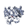
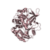
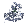
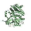
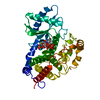
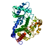
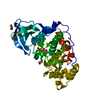
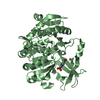
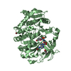
| Entry | Database: PDB / ID: 3wvn | ||||||
|---|---|---|---|---|---|---|---|
| Title | Complex structure of VinN with L-aspartate | ||||||
 Components Components | Non-ribosomal peptide synthetase | ||||||
 Keywords Keywords | LIGASE / Five-layered alpha-beta-alpha-beta-alpha sandwich fold / ATP binding | ||||||
| Function / homology |  Function and homology information Function and homology information | ||||||
| Biological species |  Streptomyces halstedii (bacteria) Streptomyces halstedii (bacteria) | ||||||
| Method |  X-RAY DIFFRACTION / SYNCHROTRON / MOLECULAR REPLACEMENT / Resolution: 2.2 Å X-RAY DIFFRACTION / SYNCHROTRON / MOLECULAR REPLACEMENT / Resolution: 2.2 Å | ||||||
 Authors Authors | Miyanaga, A. / Cieslak, J. / Shinohara, Y. / Kudo, F. / Eguchi, T. | ||||||
 Citation Citation | Journal: J.Biol.Chem. / Year: 2014 Title: The crystal structure of the adenylation enzyme VinN reveals a unique beta-amino acid recognition mechanism Authors: Miyanaga, A. / Cieslak, J. / Shinohara, Y. / Kudo, F. / Eguchi, T. | ||||||
| History |
|
- Structure visualization
| Structure viewer | Molecule: MolmilJmol/JSmol |
|---|
- Downloads & links
-Download
| PDBx/mmCIF format | 3wvn.cif.gz | 305 KB | Display | PDBx/mmCIF format |
|---|---|---|---|---|
| PDB format | pdb3wvn.ent.gz | 248 KB | Display | PDB format |
| PDBx/mmJSON format | 3wvn.json.gz | Tree view | PDBx/mmJSON format | |
| Others |  Other downloads Other downloads |
-Validation report
| Arichive directory | https://data.pdbj.org/pub/pdb/validation_reports/wv/3wvnftp://data.pdbj.org/pub/pdb/validation_reports/wv/3wvn | HTTPS FTP |
|---|
-Related structure data
| Related structure data | 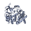 3wv4C  3wv5SC C: citing same article ( S: Starting model for refinement |
|---|---|
| Similar structure data |
-Links
 PDBj
PDBj

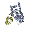
- Assembly
| Deposited unit | 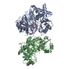
| ||||||||||||||||||
|---|---|---|---|---|---|---|---|---|---|---|---|---|---|---|---|---|---|---|---|
| 1 | 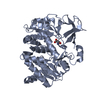
| ||||||||||||||||||
| 2 | 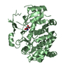
| ||||||||||||||||||
| Unit cell |
| ||||||||||||||||||
| Components on special symmetry positions |
| ||||||||||||||||||
| Noncrystallographic symmetry (NCS) | NCS domain:
NCS domain segments: Component-ID: _ / Ens-ID: 1 / Beg auth comp-ID: ASP / Beg label comp-ID: ASP / End auth comp-ID: ASP / End label comp-ID: ASP / Refine code: _ / Auth seq-ID: 24 - 601 / Label seq-ID: 40
|
-Components
| #1: Protein | Mass: 58911.902 Da / Num. of mol.: 2 Source method: isolated from a genetically manipulated source Source: (gene. exp.) Streptomyces halstedii (bacteria) / Strain: HC34 / Gene: vinN / Plasmid: pCold I / Production host: References: UniProt: Q76KY2, Ligases; Forming carbon-sulfur bonds; Acid-thiol ligases #2: Chemical |   Type: L-peptide linking / Mass: 133.103 Da / Num. of mol.: 2 / Source method: obtained synthetically / Formula: C4H7NO4 Type: L-peptide linking / Mass: 133.103 Da / Num. of mol.: 2 / Source method: obtained synthetically / Formula: C4H7NO4#3: Water | ChemComp-HOH / |  Mass: 18.015 Da / Num. of mol.: 176 / Source method: isolated from a natural source / Formula: H2O Mass: 18.015 Da / Num. of mol.: 176 / Source method: isolated from a natural source / Formula: H2O |
|---|
-Experimental details
-Experiment
| Experiment | Method: X-RAY DIFFRACTION / Number of used crystals: 1 |
|---|
- Sample preparation
| Crystal | Density Matthews: 1.87 Å3/Da / Density % sol: 34.16 % |
|---|---|
| Crystal grow | Temperature: 278 K / Method: vapor diffusion, sitting drop / pH: 8.5 Details: 0.1M Tris-HCl, 0.2M sodium acetate, 30% polyethylene glycol 4000, 100mM L-aspartate, pH 8.5, VAPOR DIFFUSION, SITTING DROP, temperature 278K |
-Data collection
| Diffraction | Mean temperature: 100 K |
|---|---|
| Diffraction source | Source: SYNCHROTRON / Site: Photon Factory  / Beamline: AR-NW12A / Wavelength: 1 Å / Beamline: AR-NW12A / Wavelength: 1 Å |
| Detector | Type: ADSC QUANTUM 210r / Detector: CCD / Date: May 26, 2014 |
| Radiation | Monochromator: Numerical link type Si(111) double crystal monochromator Protocol: SINGLE WAVELENGTH / Monochromatic (M) / Laue (L): M / Scattering type: x-ray |
| Radiation wavelength | Wavelength: 1 Å / Relative weight: 1 |
| Reflection | Resolution: 2.2→42.27 Å / Num. all: 45129 / Num. obs: 43099 / % possible obs: 95.5 % / Observed criterion σ(F): 0 / Observed criterion σ(I): 0 |
| Reflection shell | Resolution: 2.2→2.32 Å / % possible all: 88.3 |
- Processing
| Software |
| ||||||||||||||||||||||||||||||||||||||||||||||||||||||||||||||||||||||||||||||||||||||||||||||||||||||||||||||||||||||||||||||||||||||||||||||||||||||||||||||||||||||||||||||||||||||
|---|---|---|---|---|---|---|---|---|---|---|---|---|---|---|---|---|---|---|---|---|---|---|---|---|---|---|---|---|---|---|---|---|---|---|---|---|---|---|---|---|---|---|---|---|---|---|---|---|---|---|---|---|---|---|---|---|---|---|---|---|---|---|---|---|---|---|---|---|---|---|---|---|---|---|---|---|---|---|---|---|---|---|---|---|---|---|---|---|---|---|---|---|---|---|---|---|---|---|---|---|---|---|---|---|---|---|---|---|---|---|---|---|---|---|---|---|---|---|---|---|---|---|---|---|---|---|---|---|---|---|---|---|---|---|---|---|---|---|---|---|---|---|---|---|---|---|---|---|---|---|---|---|---|---|---|---|---|---|---|---|---|---|---|---|---|---|---|---|---|---|---|---|---|---|---|---|---|---|---|---|---|---|---|
| Refinement | Method to determine structure: MOLECULAR REPLACEMENT Starting model: PDB Entry 3WV5 Resolution: 2.2→39.63 Å / Cor.coef. Fo:Fc: 0.946 / Cor.coef. Fo:Fc free: 0.925 / SU B: 15.158 / SU ML: 0.166 / Cross valid method: THROUGHOUT / ESU R: 0.273 / ESU R Free: 0.209 / Stereochemistry target values: MAXIMUM LIKELIHOOD / Details: HYDROGENS HAVE BEEN ADDED IN THE RIDING POSITIONS
| ||||||||||||||||||||||||||||||||||||||||||||||||||||||||||||||||||||||||||||||||||||||||||||||||||||||||||||||||||||||||||||||||||||||||||||||||||||||||||||||||||||||||||||||||||||||
| Solvent computation | Ion probe radii: 0.8 Å / Shrinkage radii: 0.8 Å / VDW probe radii: 1.2 Å / Solvent model: MASK | ||||||||||||||||||||||||||||||||||||||||||||||||||||||||||||||||||||||||||||||||||||||||||||||||||||||||||||||||||||||||||||||||||||||||||||||||||||||||||||||||||||||||||||||||||||||
| Displacement parameters | Biso mean: 45.554 Å2
| ||||||||||||||||||||||||||||||||||||||||||||||||||||||||||||||||||||||||||||||||||||||||||||||||||||||||||||||||||||||||||||||||||||||||||||||||||||||||||||||||||||||||||||||||||||||
| Refinement step | Cycle: LAST / Resolution: 2.2→39.63 Å
| ||||||||||||||||||||||||||||||||||||||||||||||||||||||||||||||||||||||||||||||||||||||||||||||||||||||||||||||||||||||||||||||||||||||||||||||||||||||||||||||||||||||||||||||||||||||
| Refine LS restraints |
|