 Movie
Movie Controller

[English] 日本語
 Yorodumi
- PDB-3wv0: O-glycan attached to herpes simplex virus type 1 glycoprotein gB ... -
+ Open data

- Basic information
| Entry | Database: PDB / ID: 3wv0 | |||||||||
|---|---|---|---|---|---|---|---|---|---|---|
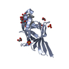
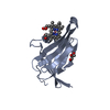
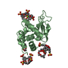
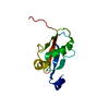
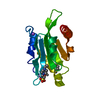
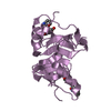
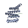
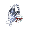
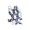
| Title | O-glycan attached to herpes simplex virus type 1 glycoprotein gB is recognized by the Ig V-set domain of human paired immunoglobulin-like type 2 receptor alpha | |||||||||
 Components Components |
| |||||||||
 Keywords Keywords | MEMBRANE PROTEIN / Immunoglobulin-like / Immunological receptor / Membrane | |||||||||
| Function / homology |  Function and homology information Function and homology informationhost cell Golgi membrane / MHC class I protein binding / host cell endosome membrane / Immunoregulatory interactions between a Lymphoid and a non-Lymphoid cell / viral envelope / symbiont entry into host cell / virion attachment to host cell / host cell plasma membrane / virion membrane / signal transduction ...host cell Golgi membrane / MHC class I protein binding / host cell endosome membrane / Immunoregulatory interactions between a Lymphoid and a non-Lymphoid cell / viral envelope / symbiont entry into host cell / virion attachment to host cell / host cell plasma membrane / virion membrane / signal transduction / extracellular exosome / identical protein binding / plasma membrane Similarity search - Function | |||||||||
| Biological species |  Homo sapiens (human) Homo sapiens (human)  Human herpesvirus 1 (Herpes simplex virus type 1) Human herpesvirus 1 (Herpes simplex virus type 1) | |||||||||
| Method |  X-RAY DIFFRACTION / SYNCHROTRON / MOLECULAR REPLACEMENT / Resolution: 2.3 Å X-RAY DIFFRACTION / SYNCHROTRON / MOLECULAR REPLACEMENT / Resolution: 2.3 Å | |||||||||
 Authors Authors | Kuroki, K. / Wang, J. / Ose, T. / Yamaguchi, M. / Tabata, S. / Maita, N. / Nakamura, S. / Kajikawa, M. / Kogure, A. / Satoh, T. ...Kuroki, K. / Wang, J. / Ose, T. / Yamaguchi, M. / Tabata, S. / Maita, N. / Nakamura, S. / Kajikawa, M. / Kogure, A. / Satoh, T. / Arase, H. / Maenaka, K. | |||||||||
 Citation Citation | Journal: Proc.Natl.Acad.Sci.USA / Year: 2014 Title: Structural basis for simultaneous recognition of an O-glycan and its attached peptide of mucin family by immune receptor PILR alpha Authors: Kuroki, K. / Wang, J. / Ose, T. / Yamaguchi, M. / Tabata, S. / Maita, N. / Nakamura, S. / Kajikawa, M. / Kogure, A. / Satoh, T. / Arase, H. / Maenaka, K. | |||||||||
| History |
|
- Structure visualization
| Structure viewer | Molecule: MolmilJmol/JSmol |
|---|
- Downloads & links
-Download
| PDBx/mmCIF format | 3wv0.cif.gz | 71 KB | Display | PDBx/mmCIF format |
|---|---|---|---|---|
| PDB format | pdb3wv0.ent.gz | 51.7 KB | Display | PDB format |
| PDBx/mmJSON format | 3wv0.json.gz | Tree view | PDBx/mmJSON format | |
| Others |  Other downloads Other downloads |
-Validation report
| Arichive directory | https://data.pdbj.org/pub/pdb/validation_reports/wv/3wv0ftp://data.pdbj.org/pub/pdb/validation_reports/wv/3wv0 | HTTPS FTP |
|---|
-Related structure data
| Related structure data | 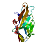 3wuzSC S: Starting model for refinement C: citing same article ( |
|---|---|
| Similar structure data |

-Links
 PDBj
PDBj- Assembly
| Deposited unit | 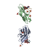
| ||||||||
|---|---|---|---|---|---|---|---|---|---|
| 1 | 
| ||||||||
| 2 | 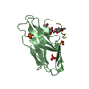
| ||||||||
| Unit cell |
|
-Components
| #1: Protein | Mass: 13967.697 Da / Num. of mol.: 2 / Fragment: V-set domain, UNP residues 32-150 / Mutation: R78G Source method: isolated from a genetically manipulated source Source: (gene. exp.) Homo sapiens (human) / Gene: PILRA / Plasmid: pGMT7 / Production host:  #2: Protein/peptide | Mass: 609.671 Da / Num. of mol.: 2 Fragment: O-glycan with attached peptide, UNP residues 50-56 Source method: obtained synthetically Details: O-glycan with peptide derived from human herpesvirus 1 Source: (synth.) Human herpesvirus 1 (Herpes simplex virus type 1)References: UniProt: P06437 #3: Polysaccharide | Source method: isolated from a genetically manipulated source #4: Chemical | ChemComp-SO4 /   Mass: 96.063 Da / Num. of mol.: 8 / Source method: obtained synthetically / Formula: SO4 Mass: 96.063 Da / Num. of mol.: 8 / Source method: obtained synthetically / Formula: SO4#5: Water | ChemComp-HOH / |  Mass: 18.015 Da / Num. of mol.: 162 / Source method: isolated from a natural source / Formula: H2O Mass: 18.015 Da / Num. of mol.: 162 / Source method: isolated from a natural source / Formula: H2OHas protein modification | Y | |
|---|
-Experimental details
-Experiment
| Experiment | Method: X-RAY DIFFRACTION / Number of used crystals: 1 |
|---|
- Sample preparation
| Crystal | Density Matthews: 2.26 Å3/Da / Density % sol: 45.69 % |
|---|---|
| Crystal grow | Temperature: 293 K / Method: vapor diffusion, hanging drop / pH: 5 Details: 0.2 M ammonium sulfate 30% PEG 8000, pH 5.0, VAPOR DIFFUSION, HANGING DROP, temperature 293K |
-Data collection
| Diffraction | Mean temperature: 100 K |
|---|---|
| Diffraction source | Source: SYNCHROTRON / Site: Photon Factory  / Beamline: BL-5A / Wavelength: 1 Å / Beamline: BL-5A / Wavelength: 1 Å |
| Detector | Type: ADSC QUANTUM 315r / Detector: CCD / Date: Jan 16, 2009 / Details: mirrors |
| Radiation | Monochromator: Si 111 / Protocol: SINGLE WAVELENGTH / Monochromatic (M) / Laue (L): M / Scattering type: x-ray |
| Radiation wavelength | Wavelength: 1 Å / Relative weight: 1 |
| Reflection | Resolution: 2.3→20 Å / Num. obs: 12122 / % possible obs: 99.7 % / Observed criterion σ(F): 0 / Observed criterion σ(I): 0 / Redundancy: 6.9 % / Biso Wilson estimate: 13.473 Å2 / Rmerge(I) obs: 0.168 / Rsym value: 0.156 / Net I/σ(I): 4.7 |
| Reflection shell | Resolution: 2.3→2.42 Å / Redundancy: 5.1 % / Rmerge(I) obs: 0.567 / Mean I/σ(I) obs: 1.5 / Rsym value: 0.512 / % possible all: 99.7 |
- Processing
| Software |
| |||||||||||||||||||||||||
|---|---|---|---|---|---|---|---|---|---|---|---|---|---|---|---|---|---|---|---|---|---|---|---|---|---|---|
| Refinement | Method to determine structure: MOLECULAR REPLACEMENT Starting model: PDB ENTRY 3WUZ Resolution: 2.3→20 Å / Isotropic thermal model: Isotropic / Cross valid method: THROUGHOUT / σ(F): 0 / σ(I): 0 / Stereochemistry target values: Engh & Huber
| |||||||||||||||||||||||||
| Displacement parameters | Biso mean: 15.141 Å2
| |||||||||||||||||||||||||
| Refinement step | Cycle: LAST / Resolution: 2.3→20 Å
| |||||||||||||||||||||||||
| Refine LS restraints |
| |||||||||||||||||||||||||
| LS refinement shell | Resolution: 2.3→2.46 Å
|
