| Entry | Database: PDB / ID: 3wjc
|
|---|
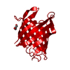
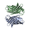
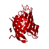
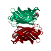
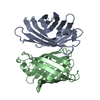
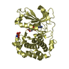
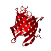
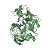
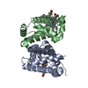
| Title | Crystal structure of mutant nitrobindin M75L/H76L/Q96C/M148L/H158L covalently linked with [Rh(Cp-Mal)(COD)] (NB4-Rh) from Arabidopsis thaliana |
|---|
 Components Components | UPF0678 fatty acid-binding protein-like protein At1g79260 |
|---|
 Keywords Keywords | TRANSPORT PROTEIN / beta-barrel / intracellular transport / hydrophobic ligands / [Rh(Cp-Mal)(COD)] |
|---|
| Function / homology |  Function and homology information Function and homology information
THAP4-like, heme-binding beta-barrel domain / Nitrobindin family / THAP4-like, heme-binding beta-barrel domain / Calycin beta-barrel core domain / Calycin / Lipocalin / Beta Barrel / Mainly BetaSimilarity search - Domain/homology |
|---|
| Biological species |   Arabidopsis thaliana (thale cress) Arabidopsis thaliana (thale cress) |
|---|
| Method |  X-RAY DIFFRACTION / MOLECULAR REPLACEMENT / Resolution: 2 Å X-RAY DIFFRACTION / MOLECULAR REPLACEMENT / Resolution: 2 Å |
|---|
 Authors Authors | Mizohata, E. / Fukumoto, K. / Onoda, A. / Bocola, M. / Arlt, M. / Inoue, T. / Schwaneberg, U. / Hayashi, T. |
|---|
 Citation Citation | Journal: CHEMCATCHEM / Year: 2014
Title: A Rhodium Complex-linked Hybrid Biocatalyst: Stereo-controlled Phenylacetylene Polymerization within an Engineered Protein Cavity
Authors: Fukumoto, K. / Onoda, A. / Mizohata, E. / Bocola, M. / Inoue, T. / Schwaneberg, U. / Hayashi, T. |
|---|
| History | | Deposition | Oct 8, 2013 | Deposition site: PDBJ / Processing site: PDBJ |
|---|
| Revision 1.0 | Apr 9, 2014 | Provider: repository / Type: Initial release |
|---|
| Revision 1.1 | Nov 6, 2024 | Group: Data collection / Database references ...Data collection / Database references / Derived calculations / Refinement description / Structure summary
Category: chem_comp_atom / chem_comp_bond ...chem_comp_atom / chem_comp_bond / database_2 / pdbx_entry_details / pdbx_modification_feature / pdbx_struct_conn_angle / struct_conn / struct_ncs_dom_lim / struct_ref_seq_dif / struct_site
Item: _database_2.pdbx_DOI / _database_2.pdbx_database_accession ..._database_2.pdbx_DOI / _database_2.pdbx_database_accession / _pdbx_struct_conn_angle.ptnr1_auth_asym_id / _pdbx_struct_conn_angle.ptnr1_auth_comp_id / _pdbx_struct_conn_angle.ptnr1_auth_seq_id / _pdbx_struct_conn_angle.ptnr1_label_asym_id / _pdbx_struct_conn_angle.ptnr1_label_atom_id / _pdbx_struct_conn_angle.ptnr1_label_comp_id / _pdbx_struct_conn_angle.ptnr1_label_seq_id / _pdbx_struct_conn_angle.ptnr2_auth_asym_id / _pdbx_struct_conn_angle.ptnr2_auth_seq_id / _pdbx_struct_conn_angle.ptnr2_label_asym_id / _pdbx_struct_conn_angle.ptnr3_auth_asym_id / _pdbx_struct_conn_angle.ptnr3_auth_comp_id / _pdbx_struct_conn_angle.ptnr3_auth_seq_id / _pdbx_struct_conn_angle.ptnr3_label_asym_id / _pdbx_struct_conn_angle.ptnr3_label_atom_id / _pdbx_struct_conn_angle.ptnr3_label_comp_id / _pdbx_struct_conn_angle.ptnr3_label_seq_id / _pdbx_struct_conn_angle.value / _struct_conn.pdbx_dist_value / _struct_conn.pdbx_leaving_atom_flag / _struct_conn.ptnr1_auth_asym_id / _struct_conn.ptnr1_auth_comp_id / _struct_conn.ptnr1_auth_seq_id / _struct_conn.ptnr1_label_asym_id / _struct_conn.ptnr1_label_atom_id / _struct_conn.ptnr1_label_comp_id / _struct_conn.ptnr1_label_seq_id / _struct_conn.ptnr2_auth_asym_id / _struct_conn.ptnr2_auth_comp_id / _struct_conn.ptnr2_auth_seq_id / _struct_conn.ptnr2_label_asym_id / _struct_conn.ptnr2_label_atom_id / _struct_conn.ptnr2_label_comp_id / _struct_ncs_dom_lim.beg_auth_comp_id / _struct_ncs_dom_lim.beg_label_asym_id / _struct_ncs_dom_lim.beg_label_comp_id / _struct_ncs_dom_lim.beg_label_seq_id / _struct_ncs_dom_lim.end_auth_comp_id / _struct_ncs_dom_lim.end_label_asym_id / _struct_ncs_dom_lim.end_label_comp_id / _struct_ncs_dom_lim.end_label_seq_id / _struct_ref_seq_dif.details / _struct_site.pdbx_auth_asym_id / _struct_site.pdbx_auth_comp_id / _struct_site.pdbx_auth_seq_id |
|---|
|
|---|
 Movie
Movie Controller

 Yorodumi
 Open data
 Basic information
 Structure visualization
 Downloads & links
 Other downloads
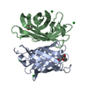


 PDBj
PDBj

 Assembly
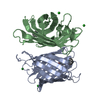

 Mass: 401.305 Da / Num. of mol.: 2 / Source method: obtained synthetically / Formula: C19H24NO2Rh
 Mass: 137.327 Da / Num. of mol.: 4 / Source method: obtained synthetically / Formula: Ba
 Mass: 18.015 Da / Num. of mol.: 174 / Source method: isolated from a natural source / Formula: H2O
 Sample preparation
 Processing