 Movie
Movie Controller

[English] 日本語
 Yorodumi
- PDB-6bg2: Crystal structure of cGMP-dependent protein kinase Ialpha (PKG Ia... -
+ Open data

- Basic information
| Entry | Database: PDB / ID: 6bg2 | ||||||
|---|---|---|---|---|---|---|---|
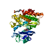
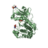
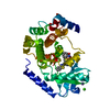
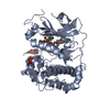
| Title | Crystal structure of cGMP-dependent protein kinase Ialpha (PKG Ialpha) catalytic domain in AMP-PNP bound state | ||||||
 Components Components | cGMP-dependent protein kinase 1 | ||||||
 Keywords Keywords | TRANSFERASE / Serine/threonine protein kinases (EC 2.7.11.12) | ||||||
| Function / homology |  Function and homology information Function and homology informationnegative regulation of inositol phosphate biosynthetic process / negative regulation of glutamate secretion / cGMP-dependent protein kinase / cGMP-dependent protein kinase activity / bone growth / cell growth involved in cardiac muscle cell development / regulation of testosterone biosynthetic process / collateral sprouting / negative regulation of platelet aggregation / positive regulation of circadian rhythm ...negative regulation of inositol phosphate biosynthetic process / negative regulation of glutamate secretion / cGMP-dependent protein kinase / cGMP-dependent protein kinase activity / bone growth / cell growth involved in cardiac muscle cell development / regulation of testosterone biosynthetic process / collateral sprouting / negative regulation of platelet aggregation / positive regulation of circadian rhythm / relaxation of vascular associated smooth muscle / Rap1 signalling / regulation of vascular permeability / : / mitogen-activated protein kinase p38 binding / cGMP effects / negative regulation of vascular associated smooth muscle cell migration / forebrain development / dendrite development / cGMP binding / negative regulation of vascular associated smooth muscle cell proliferation / spermatid development / cerebellum development / acrosomal vesicle / calcium channel regulator activity / sarcolemma / neuron migration / positive regulation of cytosolic calcium ion concentration / Ca2+ pathway / protein kinase activity / protein serine kinase activity / Golgi apparatus / signal transduction / nucleoplasm / ATP binding / identical protein binding / plasma membrane / cytoplasm / cytosol Similarity search - Function | ||||||
| Biological species |  Homo sapiens (human) Homo sapiens (human) | ||||||
| Method |  X-RAY DIFFRACTION / SYNCHROTRON / MOLECULAR REPLACEMENT / Resolution: 1.83 Å X-RAY DIFFRACTION / SYNCHROTRON / MOLECULAR REPLACEMENT / Resolution: 1.83 Å | ||||||
 Authors Authors | Qin, L. / Sankaran, B. / Kim, C. | ||||||
| Funding support |  United States, 1items United States, 1items
| ||||||
 Citation Citation | Journal: to be published Title: Crystal structure of cGMP-dependent protein kinase Ialpha (PKG Ialpha) catalytic domain Authors: Qin, L. / Sankaran, B. / Casteel, D. / Kim, C. | ||||||
| History |
|
- Structure visualization
| Structure viewer | Molecule: MolmilJmol/JSmol |
|---|
- Downloads & links
-Download
| PDBx/mmCIF format | 6bg2.cif.gz | 306.1 KB | Display | PDBx/mmCIF format |
|---|---|---|---|---|
| PDB format | pdb6bg2.ent.gz | 242.5 KB | Display | PDB format |
| PDBx/mmJSON format | 6bg2.json.gz | Tree view | PDBx/mmJSON format | |
| Others |  Other downloads Other downloads |
-Validation report
| Arichive directory | https://data.pdbj.org/pub/pdb/validation_reports/bg/6bg2ftp://data.pdbj.org/pub/pdb/validation_reports/bg/6bg2 | HTTPS FTP |
|---|
-Related structure data
| Related structure data |  6bdlC  3fjqS C: citing same article ( S: Starting model for refinement |
|---|---|
| Similar structure data |


-Links
 PDBj
PDBj


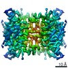
- Assembly
| Deposited unit | 
| ||||||||
|---|---|---|---|---|---|---|---|---|---|
| 1 | 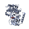
| ||||||||
| 2 | 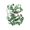
| ||||||||
| 3 | 
| ||||||||
| 4 | 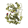
| ||||||||
| Unit cell |
|
-Components
| #1: Protein | Mass: 39473.770 Da / Num. of mol.: 4 Source method: isolated from a genetically manipulated source Details: AMP-PNP / Source: (gene. exp.) Homo sapiens (human) / Gene: PRKG1, PRKG1B, PRKGR1A, PRKGR1B / Plasmid: pBlueBacHis2A / Cell line (production host): High Five / Production host:  Trichoplusia ni (cabbage looper) / References: UniProt: Q13976, cGMP-dependent protein kinase Trichoplusia ni (cabbage looper) / References: UniProt: Q13976, cGMP-dependent protein kinase#2: Chemical | ChemComp-ANP /   Mass: 506.196 Da / Num. of mol.: 4 / Source method: obtained synthetically / Formula: C10H17N6O12P3 / Comment: AMP-PNP, energy-carrying molecule analogue*YM Mass: 506.196 Da / Num. of mol.: 4 / Source method: obtained synthetically / Formula: C10H17N6O12P3 / Comment: AMP-PNP, energy-carrying molecule analogue*YM#3: Chemical | ChemComp-MN /   Mass: 54.938 Da / Num. of mol.: 8 / Source method: obtained synthetically / Formula: Mn Mass: 54.938 Da / Num. of mol.: 8 / Source method: obtained synthetically / Formula: Mn#4: Water | ChemComp-HOH / |  Mass: 18.015 Da / Num. of mol.: 1455 / Source method: isolated from a natural source / Formula: H2O Mass: 18.015 Da / Num. of mol.: 1455 / Source method: isolated from a natural source / Formula: H2OHas protein modification | Y | |
|---|
-Experimental details
-Experiment
| Experiment | Method: X-RAY DIFFRACTION / Number of used crystals: 1 |
|---|
- Sample preparation
| Crystal | Density Matthews: 2.19 Å3/Da / Density % sol: 43.92 % |
|---|---|
| Crystal grow | Temperature: 295 K / Method: vapor diffusion, hanging drop / pH: 8 Details: 23.2% PEG1500, 0.08 M PCTP, pH8.0, 0.5% w/v polyvinylpyrrolidone K15 |
-Data collection
| Diffraction | Mean temperature: 77 K |
|---|---|
| Diffraction source | Source: SYNCHROTRON / Site: ALS / Beamline: 5.0.1 / Wavelength: 0.97741 Å |
| Detector | Type: ADSC QUANTUM 315r / Detector: CCD / Date: Sep 24, 2015 |
| Radiation | Protocol: SINGLE WAVELENGTH / Monochromatic (M) / Laue (L): M / Scattering type: x-ray |
| Radiation wavelength | Wavelength: 0.97741 Å / Relative weight: 1 |
| Reflection | Resolution: 1.83→47.23 Å / Num. obs: 117904 / % possible obs: 99 % / Redundancy: 4.7 % / CC1/2: 0.999 / Net I/σ(I): 16.8 |
| Reflection shell | Resolution: 1.83→1.86 Å / Redundancy: 4.8 % / Num. unique obs: 5795 / CC1/2: 0.698 / % possible all: 98.3 |
- Processing
| Software |
| |||||||||||||||||||||||||||||||||||||||||||||||||||||||||||||||||||||||||||||||||||||||||||||||||||||||||
|---|---|---|---|---|---|---|---|---|---|---|---|---|---|---|---|---|---|---|---|---|---|---|---|---|---|---|---|---|---|---|---|---|---|---|---|---|---|---|---|---|---|---|---|---|---|---|---|---|---|---|---|---|---|---|---|---|---|---|---|---|---|---|---|---|---|---|---|---|---|---|---|---|---|---|---|---|---|---|---|---|---|---|---|---|---|---|---|---|---|---|---|---|---|---|---|---|---|---|---|---|---|---|---|---|---|---|
| Refinement | Method to determine structure: MOLECULAR REPLACEMENT Starting model: 3FJQ Resolution: 1.83→47.231 Å / SU ML: 0.22 / Cross valid method: FREE R-VALUE / σ(F): 1.35 / Phase error: 22.86 / Stereochemistry target values: ML
| |||||||||||||||||||||||||||||||||||||||||||||||||||||||||||||||||||||||||||||||||||||||||||||||||||||||||
| Solvent computation | Shrinkage radii: 0.9 Å / VDW probe radii: 1.11 Å / Solvent model: FLAT BULK SOLVENT MODEL | |||||||||||||||||||||||||||||||||||||||||||||||||||||||||||||||||||||||||||||||||||||||||||||||||||||||||
| Refinement step | Cycle: LAST / Resolution: 1.83→47.231 Å
| |||||||||||||||||||||||||||||||||||||||||||||||||||||||||||||||||||||||||||||||||||||||||||||||||||||||||
| Refine LS restraints |
| |||||||||||||||||||||||||||||||||||||||||||||||||||||||||||||||||||||||||||||||||||||||||||||||||||||||||
| LS refinement shell |
|
