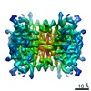
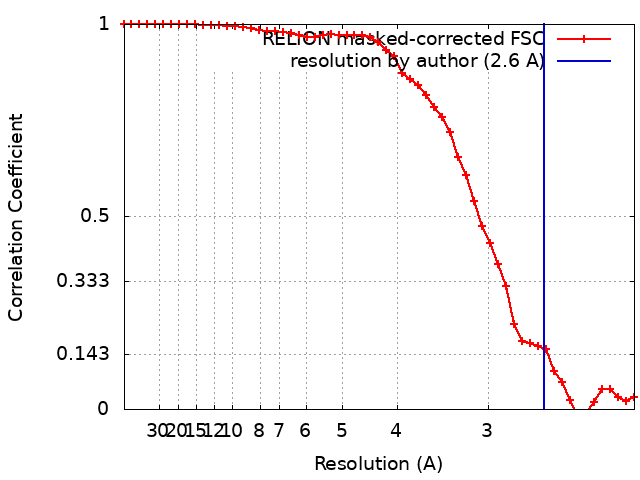
Journal: Proc Natl Acad Sci U S A / Year: 2020 Title: High-yield monolayer graphene grids for near-atomic resolution cryoelectron microscopy. Authors: Yimo Han / Xiao Fan / Haozhe Wang / Fang Zhao / Christopher G Tully / Jing Kong / Nan Yao / Nieng Yan / Abstract: Cryogenic electron microscopy (cryo-EM) has become one of the most powerful techniques to reveal the atomic structures and working mechanisms of biological macromolecules. New designs of the cryo-EM ...Cryogenic electron microscopy (cryo-EM) has become one of the most powerful techniques to reveal the atomic structures and working mechanisms of biological macromolecules. New designs of the cryo-EM grids-aimed at preserving thin, uniform vitrified ice and improving protein adsorption-have been considered a promising approach to achieving higher resolution with the minimal amount of materials and data. Here, we describe a method for preparing graphene cryo-EM grids with up to 99% monolayer graphene coverage that allows for more than 70% grid squares for effective data acquisition with improved image quality and protein density. Using our graphene grids, we have achieved 2.6-Å resolution for streptavidin, with a molecular weight of 52 kDa, from 11,000 particles. Our graphene grids increase the density of examined soluble, membrane, and lipoproteins by at least 5-fold, affording the opportunity for structural investigation of challenging proteins which cannot be produced in large quantity. In addition, our method employs only simple tools that most structural biology laboratories can access. Moreover, this approach supports customized grid designs targeting specific proteins, owing to its broad compatibility with a variety of nanomaterials.
Model: Quantifoil R1.2/1.3 / Material: GOLD / Mesh: 300 / Support film - Material: GRAPHENE / Support film - topology: CONTINUOUS / Support film - Film thickness: 0.3 nm
Vitrification
Cryogen name: ETHANE / Chamber humidity: 100 % / Chamber temperature: 281 K / Instrument: FEI VITROBOT MARK IV / Details: Blot force 0 Blot time 5s.
Details
NEB N7021S
-
Electron microscopy
Microscope
FEI TITAN KRIOS
Image recording
Film or detector model: GATAN K2 SUMMIT (4k x 4k) / Detector mode: COUNTING / Digitization - Dimensions - Width: 3838 pixel / Digitization - Dimensions - Height: 3710 pixel / Digitization - Sampling interval: 5.0 µm / Digitization - Frames/image: 1-32 / Number grids imaged: 1 / Number real images: 1086 / Average exposure time: 2.4 sec. / Average electron dose: 49.0 e/Å2
Electron beam
Acceleration voltage: 300 kV / Electron source: FIELD EMISSION GUN
Electron optics
C2 aperture diameter: 70.0 µm / Illumination mode: FLOOD BEAM / Imaging mode: BRIGHT FIELD / Cs: 0.01 mm
Experimental equipment
Model: Titan Krios / Image courtesy: FEI Company
+
Image processing
Particle selection
Number selected: 1130061 / Details: Relion auto-picking with Laplacian-of-Gaussian
In the structure databanks used in Yorodumi, some data are registered as the other names, "COVID-19 virus" and "2019-nCoV". Here are the details of the virus and the list of structure data.
Jan 31, 2019. EMDB accession codes are about to change! (news from PDBe EMDB page)
EMDB accession codes are about to change! (news from PDBe EMDB page)
The allocation of 4 digits for EMDB accession codes will soon come to an end. Whilst these codes will remain in use, new EMDB accession codes will include an additional digit and will expand incrementally as the available range of codes is exhausted. The current 4-digit format prefixed with “EMD-” (i.e. EMD-XXXX) will advance to a 5-digit format (i.e. EMD-XXXXX), and so on. It is currently estimated that the 4-digit codes will be depleted around Spring 2019, at which point the 5-digit format will come into force.
The EM Navigator/Yorodumi systems omit the EMD- prefix.
Related info.:Q: What is EMD? / ID/Accession-code notation in Yorodumi/EM Navigator
Yorodumi is a browser for structure data from EMDB, PDB, SASBDB, etc.
This page is also the successor to EM Navigator detail page, and also detail information page/front-end page for Omokage search.
The word "yorodu" (or yorozu) is an old Japanese word meaning "ten thousand". "mi" (miru) is to see.
Related info.:EMDB / PDB / SASBDB / Comparison of 3 databanks / Yorodumi Search / Aug 31, 2016. New EM Navigator & Yorodumi / Yorodumi Papers / Jmol/JSmol / Function and homology information / Changes in new EM Navigator and Yorodumi
 Movie
Movie Controller


 Open data
 Basic information
 Map data
 Sample
 Function and homology information
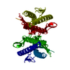
 Streptomyces avidinii (bacteria)
 Authors
 United States, 1 items
  Citation
 Structure visualization
 Downloads & links
 emd_20907.png
 http://ftp.pdbj.org/pub/emdb/structures/EMD-20907


 Z (Sec.)
 Y (Row.)
 X (Col.)


 Sample components
 Processing
 Electron microscopy
 FIELD EMISSION GUN