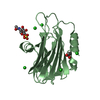
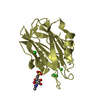
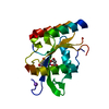
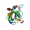
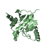
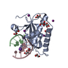
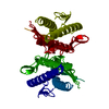
- PDB-3vp7: Crystal structure of the beta-alpha repeated, autophagy-specific ... -
+
Open data
ID or keywords:
Loading...
-
Basic information
Entry
Database: PDB / ID: 3vp7
Title
Crystal structure of the beta-alpha repeated, autophagy-specific (BARA) domain of Vps30/Atg6
Components
Vacuolar protein sorting-associated protein 30
Keywords
PROTEIN TRANSPORT / targeting / PI3-kinase complex I / pre-autophagosomal structure
Function / homology
Function and homology information
vacuole-isolation membrane contact site / cellular response to potassium ion starvation / Macroautophagy / phosphatidylinositol 3-kinase complex, class III, type II / phosphatidylinositol 3-kinase complex, class III, type I / cytoplasm to vacuole targeting by the Cvt pathway / phagophore assembly site membrane / piecemeal microautophagy of the nucleus / protein targeting to vacuole / late endosome to vacuole transport ...vacuole-isolation membrane contact site / cellular response to potassium ion starvation / Macroautophagy / phosphatidylinositol 3-kinase complex, class III, type II / phosphatidylinositol 3-kinase complex, class III, type I / cytoplasm to vacuole targeting by the Cvt pathway / phagophore assembly site membrane / piecemeal microautophagy of the nucleus / protein targeting to vacuole / late endosome to vacuole transport / phosphatidylinositol biosynthetic process / pexophagy / fungal-type vacuole membrane / retrograde transport, endosome to Golgi / cellular response to nitrogen starvation / phosphatidylinositol phosphate biosynthetic process / autophagosome assembly / mitophagy / phosphatidylinositol 3-kinase binding / macroautophagy / autophagy / protein-macromolecule adaptor activity / endosome membrane / Golgi membrane / cytosol / cytoplasm Similarity search - Function
In the structure databanks used in Yorodumi, some data are registered as the other names, "COVID-19 virus" and "2019-nCoV". Here are the details of the virus and the list of structure data.
Jan 31, 2019. EMDB accession codes are about to change! (news from PDBe EMDB page)
EMDB accession codes are about to change! (news from PDBe EMDB page)
The allocation of 4 digits for EMDB accession codes will soon come to an end. Whilst these codes will remain in use, new EMDB accession codes will include an additional digit and will expand incrementally as the available range of codes is exhausted. The current 4-digit format prefixed with “EMD-” (i.e. EMD-XXXX) will advance to a 5-digit format (i.e. EMD-XXXXX), and so on. It is currently estimated that the 4-digit codes will be depleted around Spring 2019, at which point the 5-digit format will come into force.
The EM Navigator/Yorodumi systems omit the EMD- prefix.
Related info.:Q: What is EMD? / ID/Accession-code notation in Yorodumi/EM Navigator
Yorodumi is a browser for structure data from EMDB, PDB, SASBDB, etc.
This page is also the successor to EM Navigator detail page, and also detail information page/front-end page for Omokage search.
The word "yorodu" (or yorozu) is an old Japanese word meaning "ten thousand". "mi" (miru) is to see.
Related info.:EMDB / PDB / SASBDB / Comparison of 3 databanks / Yorodumi Search / Aug 31, 2016. New EM Navigator & Yorodumi / Yorodumi Papers / Jmol/JSmol / Function and homology information / Changes in new EM Navigator and Yorodumi
 Movie
Movie Controller

 Yorodumi
 Open data
 Basic information
 Components
 Keywords
 Function and homology information
 X-RAY DIFFRACTION /
  Authors
 Citation
 Structure visualization
 Downloads & links
 Other downloads
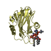


 PDBj
PDBj
 Assembly

 Mass: 18.015 Da / Num. of mol.: 42 / Source method: isolated from a natural source / Formula: H2O
 Sample preparation
 / Beamline: BL-5A / Wavelength: 0.967 Å
 Processing