 Movie
Movie Controller


+ Open data

- Basic information
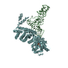
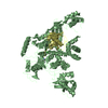
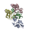
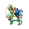
| Entry | Database: PDB / ID: 3wfs | ||||||
|---|---|---|---|---|---|---|---|
| Title | tRNA processing enzyme complex 3 | ||||||
 Components Components |
| ||||||
 Keywords Keywords | Transferase/RNA / Terminal Nucleotide Transferase / Transferase-RNA complex | ||||||
| Function / homology |  Function and homology information Function and homology informationCC tRNA cytidylyltransferase activity / tRNA surveillance / CCACCA tRNA nucleotidyltransferase activity / tRNA 3'-terminal CCA addition / Transferases; Transferring phosphorus-containing groups; Nucleotidyltransferases / tRNA binding / nucleotide binding / metal ion binding Similarity search - Function | ||||||
| Biological species | synthetic construct (others)  Aquifex aeolicus (bacteria)Thermotoga maritima MSB8 (bacteria) Aquifex aeolicus (bacteria)Thermotoga maritima MSB8 (bacteria) | ||||||
| Method |  X-RAY DIFFRACTION / SYNCHROTRON / MOLECULAR REPLACEMENT / Resolution: 3.311 Å X-RAY DIFFRACTION / SYNCHROTRON / MOLECULAR REPLACEMENT / Resolution: 3.311 Å | ||||||
 Authors Authors | Yamashita, S. / Takeshita, D. / Tomita, K. | ||||||
 Citation Citation | Journal: Structure / Year: 2014 Title: Translocation and rotation of tRNA during template-independent RNA polymerization by tRNA nucleotidyltransferase Authors: Yamashita, S. / Takeshita, D. / Tomita, K. | ||||||
| History |
|
- Structure visualization
| Structure viewer | Molecule: MolmilJmol/JSmol |
|---|
- Downloads & links
-Download
| PDBx/mmCIF format | 3wfs.cif.gz | 532.6 KB | Display | PDBx/mmCIF format |
|---|---|---|---|---|
| PDB format | pdb3wfs.ent.gz | 437.2 KB | Display | PDB format |
| PDBx/mmJSON format | 3wfs.json.gz | Tree view | PDBx/mmJSON format | |
| Others |  Other downloads Other downloads |
-Validation report
| Arichive directory | https://data.pdbj.org/pub/pdb/validation_reports/wf/3wfsftp://data.pdbj.org/pub/pdb/validation_reports/wf/3wfs | HTTPS FTP |
|---|
-Related structure data
| Related structure data | 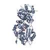 3wfoSC  3wfpC 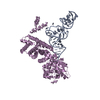 3wfqC  3wfrC 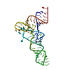 3l0uS S: Starting model for refinement C: citing same article ( |
|---|---|
| Similar structure data |


-Links
 PDBj
PDBj


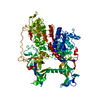
- Assembly
| Deposited unit | 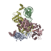
| ||||||||
|---|---|---|---|---|---|---|---|---|---|
| 1 | 
| ||||||||
| 2 | 
| ||||||||
| Unit cell |
|
-Components
| #1: RNA chain | Mass: 23850.168 Da / Num. of mol.: 2 / Source method: obtained synthetically / Source: (synth.) Thermotoga maritima MSB8 (bacteria) / References: GenBank: 498539165#2: Protein | Mass: 56943.055 Da / Num. of mol.: 2 Source method: isolated from a genetically manipulated source Source: (gene. exp.) synthetic construct (others), (gene. exp.) Aquifex aeolicus (strain VF5) (bacteria)Strain: VF5 / Gene: pcnB2 / Production host: References: UniProt: O67911, CCA tRNA nucleotidyltransferase #3: Chemical | ChemComp-SO4 /   Mass: 96.063 Da / Num. of mol.: 4 / Source method: obtained synthetically / Formula: SO4 Mass: 96.063 Da / Num. of mol.: 4 / Source method: obtained synthetically / Formula: SO4Has protein modification | N | Sequence details | THE AUTHORS CRYSTALLIZED THE ENTIRE PROTEIN, RESIDUES 1-512 FOR C, D CHAINS. THE AUTHORS KNOW THE ...THE AUTHORS CRYSTALLIZ | |
|---|
-Experimental details
-Experiment
| Experiment | Method: X-RAY DIFFRACTION / Number of used crystals: 1 |
|---|
- Sample preparation
| Crystal | Density Matthews: 3.69 Å3/Da / Density % sol: 66.63 % |
|---|
-Data collection
| Diffraction source | Source: SYNCHROTRON / Site: Photon Factory  / Beamline: BL-17A / Wavelength: 0.9791 Å / Beamline: BL-17A / Wavelength: 0.9791 Å |
|---|---|
| Detector | Date: Nov 2, 2012 |
| Radiation | Protocol: SINGLE WAVELENGTH / Monochromatic (M) / Laue (L): M / Scattering type: x-ray |
| Radiation wavelength | Wavelength: 0.9791 Å / Relative weight: 1 |
| Reflection | Resolution: 3.3→20 Å / Num. obs: 27238 / Biso Wilson estimate: 62.78 Å2 |
- Processing
| Software | Name: PHENIX / Version: (phenix.refine: 1.8.2_1309) / Classification: refinement | |||||||||||||||||||||||||||||||||||||||||||||||||||||||||||||||||||||||||||||||||||||||||||||||||||||||||||||||||||||||||||||||||||||||||||||||||||||||||||||||||||||||||||||||
|---|---|---|---|---|---|---|---|---|---|---|---|---|---|---|---|---|---|---|---|---|---|---|---|---|---|---|---|---|---|---|---|---|---|---|---|---|---|---|---|---|---|---|---|---|---|---|---|---|---|---|---|---|---|---|---|---|---|---|---|---|---|---|---|---|---|---|---|---|---|---|---|---|---|---|---|---|---|---|---|---|---|---|---|---|---|---|---|---|---|---|---|---|---|---|---|---|---|---|---|---|---|---|---|---|---|---|---|---|---|---|---|---|---|---|---|---|---|---|---|---|---|---|---|---|---|---|---|---|---|---|---|---|---|---|---|---|---|---|---|---|---|---|---|---|---|---|---|---|---|---|---|---|---|---|---|---|---|---|---|---|---|---|---|---|---|---|---|---|---|---|---|---|---|---|---|---|
| Refinement | Method to determine structure: MOLECULAR REPLACEMENT Starting model: 3WFO, 3L0U Resolution: 3.311→19.861 Å / Occupancy max: 1 / Occupancy min: 1 / FOM work R set: 0.8072 / SU ML: 0.41 / σ(F): 1.99 / Phase error: 27.1 / Stereochemistry target values: ML
| |||||||||||||||||||||||||||||||||||||||||||||||||||||||||||||||||||||||||||||||||||||||||||||||||||||||||||||||||||||||||||||||||||||||||||||||||||||||||||||||||||||||||||||||
| Solvent computation | Shrinkage radii: 0.9 Å / VDW probe radii: 1.11 Å / Solvent model: FLAT BULK SOLVENT MODEL | |||||||||||||||||||||||||||||||||||||||||||||||||||||||||||||||||||||||||||||||||||||||||||||||||||||||||||||||||||||||||||||||||||||||||||||||||||||||||||||||||||||||||||||||
| Displacement parameters | Biso max: 342.19 Å2 / Biso mean: 113.7954 Å2 / Biso min: 17.81 Å2 | |||||||||||||||||||||||||||||||||||||||||||||||||||||||||||||||||||||||||||||||||||||||||||||||||||||||||||||||||||||||||||||||||||||||||||||||||||||||||||||||||||||||||||||||
| Refinement step | Cycle: LAST / Resolution: 3.311→19.861 Å
| |||||||||||||||||||||||||||||||||||||||||||||||||||||||||||||||||||||||||||||||||||||||||||||||||||||||||||||||||||||||||||||||||||||||||||||||||||||||||||||||||||||||||||||||
| Refine LS restraints |
| |||||||||||||||||||||||||||||||||||||||||||||||||||||||||||||||||||||||||||||||||||||||||||||||||||||||||||||||||||||||||||||||||||||||||||||||||||||||||||||||||||||||||||||||
| LS refinement shell | Refine-ID: X-RAY DIFFRACTION / Total num. of bins used: 10
| |||||||||||||||||||||||||||||||||||||||||||||||||||||||||||||||||||||||||||||||||||||||||||||||||||||||||||||||||||||||||||||||||||||||||||||||||||||||||||||||||||||||||||||||
| Refinement TLS params. | Method: refined / Refine-ID: X-RAY DIFFRACTION
| |||||||||||||||||||||||||||||||||||||||||||||||||||||||||||||||||||||||||||||||||||||||||||||||||||||||||||||||||||||||||||||||||||||||||||||||||||||||||||||||||||||||||||||||
| Refinement TLS group |
|
