[English] 日本語
Yorodumi
- PDB-3w1b: Crystal Structure of Human DNA ligase IV-Artemis Complex (Mercury... -

+
Open data


ID or keywords:

Loading...

-
Basic information

Entry
Database: PDB / ID: 3w1b
Title
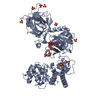
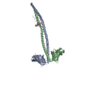
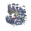
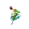
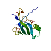
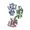
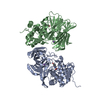
Crystal Structure of Human DNA ligase IV-Artemis Complex (Mercury Derivative)
Components
  • Artemis-derived peptide
  • DNA ligase 4
KeywordsLIGASE / DNA ligase / non-homologous end joining / DNA repair / XRCC4
Function / homology
Function and homology information


DNA ligation involved in DNA recombination / positive regulation of chromosome organization / DNA ligase IV complex / DNA ligation involved in DNA repair / DNA ligase activity / DN2 thymocyte differentiation / DNA ligase (ATP) / T cell receptor V(D)J recombination / pro-B cell differentiation / DNA ligase (ATP) activity ...DNA ligation involved in DNA recombination / positive regulation of chromosome organization / DNA ligase IV complex / DNA ligation involved in DNA repair / DNA ligase activity / DN2 thymocyte differentiation / DNA ligase (ATP) / T cell receptor V(D)J recombination / pro-B cell differentiation / DNA ligase (ATP) activity / DNA-dependent protein kinase-DNA ligase 4 complex / single strand break repair / single-stranded DNA endodeoxyribonuclease activity / immunoglobulin V(D)J recombination / nonhomologous end joining complex / 5'-3' exonuclease activity / DNA ligation / V(D)J recombination / double-strand break repair via classical nonhomologous end joining / isotype switching / 5'-3' DNA exonuclease activity / positive regulation of neurogenesis / nucleotide-excision repair, DNA gap filling / response to ionizing radiation / DNA biosynthetic process / cellular response to lithium ion / 2-LTR circle formation / ligase activity / somatic stem cell population maintenance / response to X-ray / chromosome organization / interstrand cross-link repair / condensed chromosome / telomere maintenance / B cell differentiation / neurogenesis / stem cell proliferation / central nervous system development / cellular response to ionizing radiation / response to gamma radiation / Nonhomologous End-Joining (NHEJ) / double-strand break repair via nonhomologous end joining / establishment of integrated proviral latency / positive regulation of fibroblast proliferation / double-strand break repair / fibroblast proliferation / T cell differentiation in thymus / endonuclease activity / neuron apoptotic process / adaptive immune response / in utero embryonic development / negative regulation of neuron apoptotic process / cell population proliferation / chromosome, telomeric region / damaged DNA binding / Hydrolases; Acting on ester bonds / cell cycle / cell division / intracellular membrane-bounded organelle / Golgi apparatus / magnesium ion binding / DNA binding / nucleoplasm / ATP binding / nucleus
Similarity search - Function
DNA ligase i, domain 1 / DNA ligase, ATP-dependent, N-terminal domain / DNA ligase IV domain / DNA ligase IV / DNA ligase 4 / DNA Ligase 4, adenylation domain / DNA ligase, ATP-dependent / DNA ligase, ATP-dependent, N-terminal / DNA ligase, ATP-dependent, N-terminal domain superfamily / DNA ligase N terminus ...DNA ligase i, domain 1 / DNA ligase, ATP-dependent, N-terminal domain / DNA ligase IV domain / DNA ligase IV / DNA ligase 4 / DNA Ligase 4, adenylation domain / DNA ligase, ATP-dependent / DNA ligase, ATP-dependent, N-terminal / DNA ligase, ATP-dependent, N-terminal domain superfamily / DNA ligase N terminus / DNA ligase/mRNA capping enzyme / ATP-dependent DNA ligase signature 2. / ATP-dependent DNA ligase AMP-binding site. / DNA ligase, ATP-dependent, C-terminal / ATP dependent DNA ligase C terminal region / DNA ligase, ATP-dependent, conserved site / ATP-dependent DNA ligase family profile. / DNA ligase, ATP-dependent, central / ATP dependent DNA ligase domain / DNA repair metallo-beta-lactamase / DNA repair metallo-beta-lactamase / BRCA1 C Terminus (BRCT) domain / D-amino Acid Aminotransferase; Chain A, domain 1 / breast cancer carboxy-terminal domain / BRCT domain profile. / BRCT domain / BRCT domain superfamily / Nucleic acid-binding proteins / Ribonuclease Z/Hydroxyacylglutathione hydrolase-like / OB fold (Dihydrolipoamide Acetyltransferase, E2P) / Nucleic acid-binding, OB-fold / Beta Barrel / 2-Layer Sandwich / Orthogonal Bundle / Mainly Beta / Mainly Alpha / Alpha Beta
Similarity search - Domain/homology
ADENOSINE MONOPHOSPHATE / : / DNA ligase 4 / Protein artemis
Similarity search - Component
Biological speciesHomo sapiens (human)
MethodX-RAY DIFFRACTION / SYNCHROTRON / MAD / Resolution: 2.4 Å
AuthorsOchi, T. / Blundell, T.L.
CitationJournal: Structure / Year: 2013
Title: Structure of the catalytic region of DNA ligase IV in complex with an artemis fragment sheds light on double-strand break repair
Authors: Ochi, T. / Gu, X. / Blundell, T.L.
History
DepositionNov 14, 2012Deposition site: PDBJ / Processing site: PDBJ
Revision 1.0Apr 3, 2013Provider: repository / Type: Initial release
Revision 1.1Aug 14, 2013Group: Database references

-
Structure visualization

Structure viewerMolecule:
MolmilJmol/JSmol

Downloads & links

-
Assembly

Deposited unit
A: DNA ligase 4
B: Artemis-derived peptide
hetero molecules


Theoretical massNumber of molelcules
Total (without water)74,10422
Polymers70,9912
Non-polymers3,11320
Water3,909217
1


  • Idetical with deposited unit
  • defined by author&software
TypeNameSymmetry operationNumber
identity operation1_555x,y,z1
Buried area3950 Å2
ΔGint-326 kcal/mol
Surface area27430 Å2
MethodPISA
Unit cell
Length a, b, c (Å)68.630, 105.210, 122.040
Angle α, β, γ (deg.)90.00, 90.00, 90.00
Int Tables number18
Space group name H-MP22121

-
Components

-
Protein / Protein/peptide , 2 types, 2 molecules AB

#1: Protein DNA ligase 4 / DNA ligase IV / Polydeoxyribonucleotide synthase [ATP] 4


Mass: 69538.312 Da / Num. of mol.: 1 / Fragment: Catalytic region, UNP residues 1-609
Source method: isolated from a genetically manipulated source
Details: Hippocampus / Source: (gene. exp.) Homo sapiens (human) / Gene: LIG4 / Plasmid: pOPINS / Production host: Escherichia coli (E. coli) / Strain (production host): Rosetta2(DE3)pLysS / References: UniProt: P49917, DNA ligase (ATP)
#2: Protein/peptide Artemis-derived peptide / DNA cross-link repair 1C protein / Protein A-SCID / SNM1 homolog C / hSNM1C / SNM1-like protein


Mass: 1452.653 Da / Num. of mol.: 1 / Source method: obtained synthetically / Source: (synth.) Homo sapiens (human) / References: UniProt: Q96SD1

-
Non-polymers , 4 types, 237 molecules

#3: Chemical ChemComp-AMP / ADENOSINE MONOPHOSPHATE


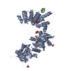
Mass: 347.221 Da / Num. of mol.: 1 / Source method: obtained synthetically / Formula: C10H14N5O7P / Comment: AMP*YM
#4: Chemical
ChemComp-SO4 / SULFATE ION


Mass: 96.063 Da / Num. of mol.: 10 / Source method: obtained synthetically / Formula: SO4
#5: Chemical
ChemComp-HG / MERCURY (II) ION


Mass: 200.590 Da / Num. of mol.: 9 / Source method: obtained synthetically / Formula: Hg
#6: Water ChemComp-HOH / water


Mass: 18.015 Da / Num. of mol.: 217 / Source method: isolated from a natural source / Formula: H2O

-
Experimental details

-
Experiment

ExperimentMethod: X-RAY DIFFRACTION / Number of used crystals: 1

-
Sample preparation

CrystalDensity Matthews: 3.1 Å3/Da / Density % sol: 60.36 %
Crystal growTemperature: 291 K / Method: vapor diffusion, hanging drop / pH: 5.6
Details: 2M ammonium sulfate, 10mM YCl, 100mM MES, pH 5.6, VAPOR DIFFUSION, HANGING DROP, temperature 291K

-
Data collection

DiffractionMean temperature: 100 K
Diffraction sourceSource: SYNCHROTRON / Site: Diamond / Beamline: I04 / Wavelength: 1.0036, 1.0093, 0.9915
DetectorType: ADSC QUANTUM 315r / Detector: CCD / Date: Oct 10, 2012
RadiationMonochromator: Double crystal / Protocol: MAD / Monochromatic (M) / Laue (L): M / Scattering type: x-ray
Radiation wavelength
IDWavelength (Å)Relative weight
11.00361
21.00931
30.99151
ReflectionResolution: 2.4→59.82 Å / Num. all: 35180 / Num. obs: 34931 / % possible obs: 99.3 % / Observed criterion σ(I): 2 / Redundancy: 3.5 % / Biso Wilson estimate: 38.14 Å2 / Rmerge(I) obs: 0.087 / Net I/σ(I): 8.5
Reflection shellResolution: 2.4→2.53 Å / Redundancy: 3.6 % / Rmerge(I) obs: 0.536 / Mean I/σ(I) obs: 1.9 / Num. measured obs: 5069 / % possible all: 100

-
Processing

Software
NameVersionClassification
DNAdata collection
SOLVEphasing
PHENIX(phenix.refine: 1.8.1_1168)refinement
MOSFLMdata reduction
SCALAdata scaling
RefinementMethod to determine structure: MAD / Resolution: 2.4→59.82 Å / Occupancy max: 1 / Occupancy min: 0.4 / SU ML: 0.29 / σ(F): 1.35 / Phase error: 21.75 / Stereochemistry target values: ML
RfactorNum. reflection% reflection
Rfree0.2253 1994 5.71 %
Rwork0.1763 --
obs0.1791 34921 98.93 %
all-35298 -
Solvent computationShrinkage radii: 0.9 Å / VDW probe radii: 1.11 Å / Solvent model: FLAT BULK SOLVENT MODEL
Displacement parametersBiso max: 174.78 Å2 / Biso mean: 43.5842 Å2 / Biso min: 15.38 Å2
Refinement stepCycle: LAST / Resolution: 2.4→59.82 Å
ProteinNucleic acidLigandSolventTotal
Num. atoms4723 0 63 217 5003
Refine LS restraints
Refine-IDTypeDev idealNumber
X-RAY DIFFRACTIONf_bond_d0.0094868
X-RAY DIFFRACTIONf_angle_d1.1376580
X-RAY DIFFRACTIONf_dihedral_angle_d15.2461797
X-RAY DIFFRACTIONf_chiral_restr0.078726
X-RAY DIFFRACTIONf_plane_restr0.005832
LS refinement shell

Refine-ID: X-RAY DIFFRACTION / Total num. of bins used: 14

Resolution (Å)Rfactor RfreeNum. reflection RfreeRfactor RworkNum. reflection RworkNum. reflection all% reflection obs (%)
2.4-2.460.31461430.249723502493100
2.46-2.52650.32421400.235623082448100
2.5265-2.60090.30371430.229423512494100
2.6009-2.68480.29571410.221623352476100
2.6848-2.78080.26611430.213223522495100
2.7808-2.89210.28061420.202223322474100
2.8921-3.02380.2681420.223482490100
3.0238-3.18320.25541420.1962337247999
3.1832-3.38260.22031430.17252350249399
3.3826-3.64370.21261430.15922348249199
3.6437-4.01030.21351420.15132356249899
4.0103-4.59050.17391440.13292364250898
4.5905-5.78280.18041400.15292350249096
5.7828-59.83850.19941460.18012446259296
Refinement TLS params.

Method: refined / Refine-ID: X-RAY DIFFRACTION

IDL112)L122)L132)L222)L232)L332)S11 (Å °)S12 (Å °)S13 (Å °)S21 (Å °)S22 (Å °)S23 (Å °)S31 (Å °)S32 (Å °)S33 (Å °)T112)T122)T132)T222)T232)T332)Origin x (Å)Origin y (Å)Origin z (Å)
11.8592-1.01-0.19542.6799-0.18012.35820.01260.047-0.04220.02560.08880.01480.0436-0.0812-0.09760.1883-0.01670.00310.2128-0.01470.1806-9.562224.48453.6488
21.69610.0888-0.45212.13441.3084.56120.14630.033-0.0037-0.0783-0.03540.1569-0.4159-0.625-0.09740.31070.07680.0390.35650.1050.2725-19.050639.799722.4208
35.41620.34290.06931.50390.43242.79960.1503-0.1728-0.6615-0.0132-0.03640.10970.2993-0.1946-0.09050.2579-0.0028-0.0760.27830.04850.4352-21.055317.197-2.3832
48.503-6.6887-3.45885.34862.99042.5231-0.2229-0.57310.33191.31360.1606-0.61430.6278-0.57340.09860.65370.01630.06840.6405-0.00510.4345-11.878331.141772.053
51.296-0.4633-0.67520.80790.6050.5291-0.0573-0.14230.1658-0.1634-0.04420.1680.1731-0.02830.10550.6999-0.005-0.0510.82020.09630.6398-11.486331.551231.3755
6-0.1810.0326-0.067-0.0840.13960.29980.04150.0396-0.06050.00590.02240.0565-0.088-0.0981-0.06250.25410.05630.04080.310.02190.2904-14.129528.818534.2651
7-0.22330.06980.43361.62550.16240.379-0.1761-0.1084-0.2778-0.310.14160.2252-0.3411-0.18530.02780.49440.00820.1290.9738-0.02780.7427-14.647723.870612.541
Refinement TLS group
IDRefine-IDRefine TLS-IDSelection detailsAuth asym-IDAuth seq-ID
1X-RAY DIFFRACTION1chain A and resseq 6:241A6 - 241
2X-RAY DIFFRACTION2chain A and resseq 242:454 or chain A and resseq 701:701A242 - 454
3X-RAY DIFFRACTION2chain A and resseq 242:454 or chain A and resseq 701:701A701
4X-RAY DIFFRACTION3chain A and resseq 455:604A455 - 605
5X-RAY DIFFRACTION4chain B and resseq 486:495B486 - 495
6X-RAY DIFFRACTION5chain A and resseq 702:711A702 - 711
7X-RAY DIFFRACTION6chain A and resseq 801:1016 or chain B and resseq 501:501A801 - 1016
8X-RAY DIFFRACTION6chain A and resseq 801:1016 or chain B and resseq 501:501B501
9X-RAY DIFFRACTION7chain A and resseq 712:720A712 - 720

+
About Yorodumi

-
News

-
Feb 9, 2022. New format data for meta-information of EMDB entries

New format data for meta-information of EMDB entries

  • Version 3 of the EMDB header file is now the official format.
  • The previous official version 1.9 will be removed from the archive.

Related info.:EMDB header

External links:wwPDB to switch to version 3 of the EMDB data model

-
Aug 12, 2020. Covid-19 info

Covid-19 info

URL: https://pdbjlvh1.pdbj.org/emnavi/covid19.php

New page: Covid-19 featured information page in EM Navigator.

Related info.:Covid-19 info / Mar 5, 2020. Novel coronavirus structure data

+
Mar 5, 2020. Novel coronavirus structure data

Novel coronavirus structure data

Related info.:Yorodumi Speices / Aug 12, 2020. Covid-19 info

External links:COVID-19 featured content - PDBj / Molecule of the Month (242):Coronavirus Proteases

+
Jan 31, 2019. EMDB accession codes are about to change! (news from PDBe EMDB page)

EMDB accession codes are about to change! (news from PDBe EMDB page)

  • The allocation of 4 digits for EMDB accession codes will soon come to an end. Whilst these codes will remain in use, new EMDB accession codes will include an additional digit and will expand incrementally as the available range of codes is exhausted. The current 4-digit format prefixed with “EMD-” (i.e. EMD-XXXX) will advance to a 5-digit format (i.e. EMD-XXXXX), and so on. It is currently estimated that the 4-digit codes will be depleted around Spring 2019, at which point the 5-digit format will come into force.
  • The EM Navigator/Yorodumi systems omit the EMD- prefix.

Related info.:Q: What is EMD? / ID/Accession-code notation in Yorodumi/EM Navigator

External links:EMDB Accession Codes are Changing Soon! / Contact to PDBj

+
Jul 12, 2017. Major update of PDB

Major update of PDB

  • wwPDB released updated PDB data conforming to the new PDBx/mmCIF dictionary.
  • This is a major update changing the version number from 4 to 5, and with Remediation, in which all the entries are updated.
  • In this update, many items about electron microscopy experimental information are reorganized (e.g. em_software).
  • Now, EM Navigator and Yorodumi are based on the updated data.

External links:wwPDB Remediation / Enriched Model Files Conforming to OneDep Data Standards Now Available in the PDB FTP Archive

-
Yorodumi

Thousand views of thousand structures

  • Yorodumi is a browser for structure data from EMDB, PDB, SASBDB, etc.
  • This page is also the successor to EM Navigator detail page, and also detail information page/front-end page for Omokage search.
  • The word "yorodu" (or yorozu) is an old Japanese word meaning "ten thousand". "mi" (miru) is to see.

Related info.:EMDB / PDB / SASBDB / Comparison of 3 databanks / Yorodumi Search / Aug 31, 2016. New EM Navigator & Yorodumi / Yorodumi Papers / Jmol/JSmol / Function and homology information / Changes in new EM Navigator and Yorodumi

Read more