- PDB-1x9n: Crystal Structure of Human DNA Ligase I bound to 5'-adenylated, n... -
+
Open data
ID or keywords:
Loading...
-
Basic information
Entry
Database: PDB / ID: 1x9n
Title
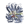
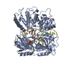
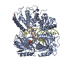
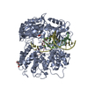
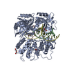
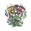
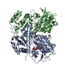
Crystal Structure of Human DNA Ligase I bound to 5'-adenylated, nicked DNA
Components
5'-phosphorylated DNA
DNA ligase I
dideoxy terminated DNA
template DNA
Keywords
Ligase/DNA / DNA ligase / 5'-adenylated nicked DNA / protein-DNA complex / Ligase-DNA COMPLEX
Function / homology
Function and homology information
Okazaki fragment processing involved in mitotic DNA replication / DNA ligase activity / DNA ligase (ATP) / DNA ligase (ATP) activity / telomere maintenance via semi-conservative replication / Regulation of MITF-M-dependent genes involved in DNA replication, damage repair and senescence / Processive synthesis on the lagging strand / lagging strand elongation / Mismatch repair (MMR) directed by MSH2:MSH3 (MutSbeta) / Mismatch repair (MMR) directed by MSH2:MSH6 (MutSalpha) ...Okazaki fragment processing involved in mitotic DNA replication / DNA ligase activity / DNA ligase (ATP) / DNA ligase (ATP) activity / telomere maintenance via semi-conservative replication / Regulation of MITF-M-dependent genes involved in DNA replication, damage repair and senescence / Processive synthesis on the lagging strand / lagging strand elongation / Mismatch repair (MMR) directed by MSH2:MSH3 (MutSbeta) / Mismatch repair (MMR) directed by MSH2:MSH6 (MutSalpha) / Processive synthesis on the C-strand of the telomere / DNA biosynthetic process / Early Phase of HIV Life Cycle / POLB-Dependent Long Patch Base Excision Repair / PCNA-Dependent Long Patch Base Excision Repair / anatomical structure morphogenesis / mismatch repair / base-excision repair, gap-filling / Gap-filling DNA repair synthesis and ligation in GG-NER / base-excision repair / Gap-filling DNA repair synthesis and ligation in TC-NER / DNA recombination / cell division / DNA repair / DNA binding / nucleoplasm / ATP binding / metal ion binding / nucleus Similarity search - Function
DNA ligase i, domain 1 / DNA ligase, ATP-dependent, N-terminal domain / Dna Ligase; domain 1 - #70 / : / DNA ligase/mRNA capping enzyme / DNA ligase, ATP-dependent / DNA ligase, ATP-dependent, N-terminal / DNA ligase, ATP-dependent, N-terminal domain superfamily / DNA ligase N terminus / ATP-dependent DNA ligase AMP-binding site. ...DNA ligase i, domain 1 / DNA ligase, ATP-dependent, N-terminal domain / Dna Ligase; domain 1 - #70 / : / DNA ligase/mRNA capping enzyme / DNA ligase, ATP-dependent / DNA ligase, ATP-dependent, N-terminal / DNA ligase, ATP-dependent, N-terminal domain superfamily / DNA ligase N terminus / ATP-dependent DNA ligase AMP-binding site. / ATP-dependent DNA ligase signature 2. / DNA ligase, ATP-dependent, C-terminal / ATP dependent DNA ligase C terminal region / DNA ligase, ATP-dependent, conserved site / ATP-dependent DNA ligase family profile. / DNA ligase, ATP-dependent, central / ATP dependent DNA ligase domain / D-amino Acid Aminotransferase; Chain A, domain 1 / Nucleic acid-binding proteins / Dna Ligase; domain 1 / OB fold (Dihydrolipoamide Acetyltransferase, E2P) / Nucleic acid-binding, OB-fold / Beta Barrel / 2-Layer Sandwich / Orthogonal Bundle / Mainly Beta / Mainly Alpha / Alpha Beta Similarity search - Domain/homology
In the structure databanks used in Yorodumi, some data are registered as the other names, "COVID-19 virus" and "2019-nCoV". Here are the details of the virus and the list of structure data.
Jan 31, 2019. EMDB accession codes are about to change! (news from PDBe EMDB page)
EMDB accession codes are about to change! (news from PDBe EMDB page)
The allocation of 4 digits for EMDB accession codes will soon come to an end. Whilst these codes will remain in use, new EMDB accession codes will include an additional digit and will expand incrementally as the available range of codes is exhausted. The current 4-digit format prefixed with “EMD-” (i.e. EMD-XXXX) will advance to a 5-digit format (i.e. EMD-XXXXX), and so on. It is currently estimated that the 4-digit codes will be depleted around Spring 2019, at which point the 5-digit format will come into force.
The EM Navigator/Yorodumi systems omit the EMD- prefix.
Related info.:Q: What is EMD? / ID/Accession-code notation in Yorodumi/EM Navigator
Yorodumi is a browser for structure data from EMDB, PDB, SASBDB, etc.
This page is also the successor to EM Navigator detail page, and also detail information page/front-end page for Omokage search.
The word "yorodu" (or yorozu) is an old Japanese word meaning "ten thousand". "mi" (miru) is to see.
Related info.:EMDB / PDB / SASBDB / Comparison of 3 databanks / Yorodumi Search / Aug 31, 2016. New EM Navigator & Yorodumi / Yorodumi Papers / Jmol/JSmol / Function and homology information / Changes in new EM Navigator and Yorodumi
 Movie
Movie Controller

 Yorodumi
 Open data
 Basic information
 Components
 Keywords
 Function and homology information
 Homo sapiens (human)
 X-RAY DIFFRACTION /
  Authors
 Citation
 Structure visualization
 Downloads & links
 Other downloads
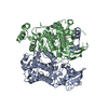


 PDBj
PDBj


 Assembly


 Mass: 347.221 Da / Num. of mol.: 1 / Source method: obtained synthetically / Formula: C10H14N5O7P / Comment: AMP*YM
 Sample preparation
 Processing