 Movie
Movie Controller

[English] 日本語
 Yorodumi
- PDB-1ehi: D-ALANINE:D-LACTATE LIGASE (LMDDL2) OF VANCOMYCIN-RESISTANT LEUCO... -
+ Open data

- Basic information
| Entry | Database: PDB / ID: 1ehi | ||||||
|---|---|---|---|---|---|---|---|
| Title | D-ALANINE:D-LACTATE LIGASE (LMDDL2) OF VANCOMYCIN-RESISTANT LEUCONOSTOC MESENTEROIDES | ||||||
 Components Components | D-ALANINE:D-LACTATE LIGASE | ||||||
 Keywords Keywords | LIGASE / ATP-binding. Grasp motif for ATP. | ||||||
| Function / homology |  Function and homology information Function and homology informationD-alanine-D-alanine ligase / D-alanine-D-alanine ligase activity / peptidoglycan biosynthetic process / cell wall organization / regulation of cell shape / ATP binding / metal ion binding / cytosol Similarity search - Function | ||||||
| Biological species |  Leuconostoc mesenteroides (bacteria) Leuconostoc mesenteroides (bacteria) | ||||||
| Method |  X-RAY DIFFRACTION / SYNCHROTRON / 3-wavelength MAD near Se edge (x12c at BNL) / Resolution: 2.38 Å X-RAY DIFFRACTION / SYNCHROTRON / 3-wavelength MAD near Se edge (x12c at BNL) / Resolution: 2.38 Å | ||||||
 Authors Authors | Kuzin, A.P. / Sun, T. / Jorczak-Baillass, J. / Healy, V.L. / Walsh, C.T. / Knox, J.R. | ||||||
 Citation Citation | Journal: Structure Fold.Des. / Year: 2000 Title: Enzymes of vancomycin resistance: the structure of D-alanine-D-lactate ligase of naturally resistant Leuconostoc mesenteroides. Authors: Kuzin, A.P. / Sun, T. / Jorczak-Baillass, J. / Healy, V.L. / Walsh, C.T. / Knox, J.R. #1: Journal: Science / Year: 1994Title: Vancomycin Resistance: Structure of D-alanine:D-alanine Ligase at 2.3 A Resolution Authors: Fan, C. / Moews, P.C. / Walsh, C.T. / Knox, J.R. #2: Journal: Biochemistry / Year: 1997Title: D-alanine:D-alanine ligase: Phosphonate and Phosphinate Intermediates with Wild-type and the Y216F Mutant Authors: Fan, C. / Park, I.-S. / Walsh, C.T. / Knox, J.R. | ||||||
| History |
|
- Structure visualization
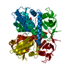
| Structure viewer | Molecule: MolmilJmol/JSmol |
|---|
- Downloads & links
-Download
| PDBx/mmCIF format | 1ehi.cif.gz | 156.8 KB | Display | PDBx/mmCIF format |
|---|---|---|---|---|
| PDB format | pdb1ehi.ent.gz | 122.4 KB | Display | PDB format |
| PDBx/mmJSON format | 1ehi.json.gz | Tree view | PDBx/mmJSON format | |
| Others |  Other downloads Other downloads |
-Validation report
| Arichive directory | https://data.pdbj.org/pub/pdb/validation_reports/eh/1ehiftp://data.pdbj.org/pub/pdb/validation_reports/eh/1ehi | HTTPS FTP |
|---|
-Related structure data
| Related structure data | |
|---|---|
| Similar structure data |


-Links
 PDBj
PDBj


- Assembly
| Deposited unit | 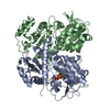
| ||||||||
|---|---|---|---|---|---|---|---|---|---|
| 1 |
| ||||||||
| Unit cell |
| ||||||||
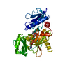
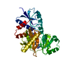
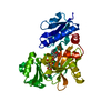
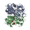
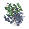
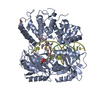
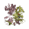
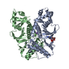
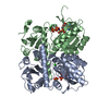
| Details | The ligase is a dimer formed of chain A and B. However, part of chain B is missing in the crystallographic model. |
-Components
| #1: Protein | Mass: 41866.883 Da / Num. of mol.: 2 Source method: isolated from a genetically manipulated source Source: (gene. exp.) Leuconostoc mesenteroides (bacteria) / Plasmid: PLMDDL2 / Production host: References: UniProt: P71454, UniProt: Q03ZI1*PLUS, D-alanine-D-alanine ligase #2: Chemical |   Mass: 24.305 Da / Num. of mol.: 2 / Source method: obtained synthetically / Formula: Mg Mass: 24.305 Da / Num. of mol.: 2 / Source method: obtained synthetically / Formula: MgDetails: Made by Prof. Paul A. Bartlett, Dept. of Chem., UC Berkeley #3: Chemical | ChemComp-ADP / |   Mass: 427.201 Da / Num. of mol.: 1 / Source method: obtained synthetically / Formula: C10H15N5O10P2 / Comment: ADP, energy-carrying molecule*YM Mass: 427.201 Da / Num. of mol.: 1 / Source method: obtained synthetically / Formula: C10H15N5O10P2 / Comment: ADP, energy-carrying molecule*YM#4: Chemical | ChemComp-PHY / | 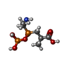  Mass: 275.133 Da / Num. of mol.: 1 / Source method: obtained synthetically / Formula: C6H15NO7P2 Mass: 275.133 Da / Num. of mol.: 1 / Source method: obtained synthetically / Formula: C6H15NO7P2#5: Water | ChemComp-HOH / |  Mass: 18.015 Da / Num. of mol.: 213 / Source method: isolated from a natural source / Formula: H2O Mass: 18.015 Da / Num. of mol.: 213 / Source method: isolated from a natural source / Formula: H2O |
|---|
-Experimental details
-Experiment
| Experiment | Method: X-RAY DIFFRACTION / Number of used crystals: 1 |
|---|
- Sample preparation
| Crystal | Density Matthews: 2.6 Å3/Da / Density % sol: 53.2 % | ||||||||||||||||||||||||||||||||||||||||||||||||||||||||||||||||||||||||||||||||||||
|---|---|---|---|---|---|---|---|---|---|---|---|---|---|---|---|---|---|---|---|---|---|---|---|---|---|---|---|---|---|---|---|---|---|---|---|---|---|---|---|---|---|---|---|---|---|---|---|---|---|---|---|---|---|---|---|---|---|---|---|---|---|---|---|---|---|---|---|---|---|---|---|---|---|---|---|---|---|---|---|---|---|---|---|---|---|
| Crystal grow | Method: vapor diffusion, sitting drop / pH: 6.5 Details: PEG8000 15%, 0.1 M ammonium sulfate, 50mM NaCl, 2.4mM DTT, 1mM MgCl2, 3mM ATP, 3mM phosphinic acid, 50mM MES buffer., pH 6.5, VAPOR DIFFUSION, SITTING DROP Temp details: 293-295 | ||||||||||||||||||||||||||||||||||||||||||||||||||||||||||||||||||||||||||||||||||||
| Crystal grow | *PLUS Method: vapor diffusion | ||||||||||||||||||||||||||||||||||||||||||||||||||||||||||||||||||||||||||||||||||||
| Components of the solutions | *PLUS
|
-Data collection
| Diffraction | Mean temperature: 100 K |
|---|---|
| Diffraction source | Source: SYNCHROTRON / Site: CHESS  / Beamline: A1 / Wavelength: 0.908 / Beamline: A1 / Wavelength: 0.908 |
| Detector | Type: ADSC / Detector: CCD / Date: Aug 15, 1997 |
| Radiation | Protocol: MAD / Monochromatic (M) / Laue (L): M / Scattering type: x-ray |
| Radiation wavelength | Wavelength: 0.908 Å / Relative weight: 1 |
| Reflection | Resolution: 2.38→100 Å / Num. all: 30564 / Num. obs: 30564 / % possible obs: 82 % / Observed criterion σ(F): 0 / Observed criterion σ(I): 0 / Redundancy: 3.1 % / Rmerge(I) obs: 0.072 / Net I/σ(I): 11 |
| Reflection shell | Resolution: 2.38→2.48 Å / Redundancy: 2.5 % / Rmerge(I) obs: 0.21 / Mean I/σ(I) obs: 3.8 / Num. unique all: 1738 / % possible all: 47 |
| Reflection | *PLUS % possible obs: 82 % / Num. measured all: 94338 |
| Reflection shell | *PLUS % possible obs: 47 % / Num. unique obs: 1738 / Num. measured obs: 4374 |
- Processing
| Software |
| |||||||||||||||||||||||||
|---|---|---|---|---|---|---|---|---|---|---|---|---|---|---|---|---|---|---|---|---|---|---|---|---|---|---|
| Refinement | Method to determine structure: 3-wavelength MAD near Se edge (x12c at BNL) Resolution: 2.38→100 Å / σ(F): 0 / σ(I): 0 / Stereochemistry target values: Engh & Huber Details: bulk solvent correction. A 3-wavelength MAD method was used to determine structure of the selenomet-protein. A native data set (A1 at CHESS) was used to refine structure.
| |||||||||||||||||||||||||
| Refinement step | Cycle: LAST / Resolution: 2.38→100 Å
| |||||||||||||||||||||||||
| Refine LS restraints |
| |||||||||||||||||||||||||
| Software | *PLUS Name: X-PLOR / Version: 3.851 / Classification: refinement | |||||||||||||||||||||||||
| Refinement | *PLUS Lowest resolution: 100 Å / σ(F): 0 | |||||||||||||||||||||||||
| Solvent computation | *PLUS | |||||||||||||||||||||||||
| Displacement parameters | *PLUS | |||||||||||||||||||||||||
| Refine LS restraints | *PLUS
|
