 Movie
Movie Controller

[English] 日本語
 Yorodumi
- PDB-4me6: Crystal structure of D-alanine-D-alanine ligase A from Xanthomona... -
+ Open data

- Basic information
| Entry | Database: PDB / ID: 4me6 | ||||||
|---|---|---|---|---|---|---|---|
| Title | Crystal structure of D-alanine-D-alanine ligase A from Xanthomonas oryzae pathovar oryzae with ADP | ||||||
 Components Components | D-alanine--D-alanine ligase | ||||||
 Keywords Keywords | LIGASE / ADP-depending enzyme / d-alanine-d-alanine ligase / Nucleotide binding | ||||||
| Function / homology |  Function and homology information Function and homology informationD-alanine-D-alanine ligase / D-alanine-D-alanine ligase activity / peptidoglycan biosynthetic process / cell wall organization / regulation of cell shape / ATP binding / metal ion binding / cytosol Similarity search - Function | ||||||
| Biological species |  Xanthomonas oryzae pv. oryzae (bacteria) Xanthomonas oryzae pv. oryzae (bacteria) | ||||||
| Method |  X-RAY DIFFRACTION / SYNCHROTRON / MOLECULAR REPLACEMENT / Resolution: 2.1 Å X-RAY DIFFRACTION / SYNCHROTRON / MOLECULAR REPLACEMENT / Resolution: 2.1 Å | ||||||
 Authors Authors | Doan, T.T.N. / Kim, J.K. / Ahn, Y.J. / Lee, B.M. / Kang, L.W. | ||||||
 Citation Citation | Journal: Arch.Biochem.Biophys. / Year: 2014 Title: Crystal structures of d-alanine-d-alanine ligase from Xanthomonas oryzae pv. oryzae alone and in complex with nucleotides Authors: Doan, T.N.N. / Kim, J.K. / Ngo, H.P.T. / Tran, H.T. / Cha, S.S. / Chung, K.M. / Huynh, K.H. / Ahn, Y.J. / Kang, L.W. | ||||||
| History |
|
- Structure visualization
| Structure viewer | Molecule: MolmilJmol/JSmol |
|---|
- Downloads & links
-Download
| PDBx/mmCIF format | 4me6.cif.gz | 85.4 KB | Display | PDBx/mmCIF format |
|---|---|---|---|---|
| PDB format | pdb4me6.ent.gz | 61.1 KB | Display | PDB format |
| PDBx/mmJSON format | 4me6.json.gz | Tree view | PDBx/mmJSON format | |
| Others |  Other downloads Other downloads |
-Validation report
| Arichive directory | https://data.pdbj.org/pub/pdb/validation_reports/me/4me6ftp://data.pdbj.org/pub/pdb/validation_reports/me/4me6 | HTTPS FTP |
|---|
-Related structure data
| Related structure data |  4l1kC  3e5nS S: Starting model for refinement C: citing same article ( |
|---|---|
| Similar structure data |


-Links
 PDBj
PDBj


- Assembly
| Deposited unit | 
| ||||||||
|---|---|---|---|---|---|---|---|---|---|
| 1 | 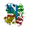
| ||||||||
| Unit cell |
|
-Components
| #1: Protein | Mass: 41945.777 Da / Num. of mol.: 1 / Mutation: M327V Source method: isolated from a genetically manipulated source Source: (gene. exp.) Xanthomonas oryzae pv. oryzae (bacteria)Strain: KACC10331 / Gene: ddlA, ddl, XOO0352 / Plasmid: pET11a / Production host: |
|---|---|
| #2: Chemical | ChemComp-MG /   Mass: 24.305 Da / Num. of mol.: 1 / Source method: obtained synthetically / Formula: Mg Mass: 24.305 Da / Num. of mol.: 1 / Source method: obtained synthetically / Formula: Mg |
| #3: Chemical | ChemComp-ADP /   Mass: 427.201 Da / Num. of mol.: 1 / Source method: obtained synthetically / Formula: C10H15N5O10P2 / Comment: ADP, energy-carrying molecule*YM Mass: 427.201 Da / Num. of mol.: 1 / Source method: obtained synthetically / Formula: C10H15N5O10P2 / Comment: ADP, energy-carrying molecule*YM |
| #4: Water | ChemComp-HOH /  Mass: 18.015 Da / Num. of mol.: 171 / Source method: isolated from a natural source / Formula: H2O Mass: 18.015 Da / Num. of mol.: 171 / Source method: isolated from a natural source / Formula: H2O |
| Sequence details | RESIDUES 1-21 ARE PART OF THE MATURE PROTEIN ACCORDING TO DATABASE GI:122879012 (PROTEIN REF YP_198991.6). |
-Experimental details
-Experiment
| Experiment | Method: X-RAY DIFFRACTION / Number of used crystals: 1 |
|---|
- Sample preparation
| Crystal | Density Matthews: 2.03 Å3/Da / Density % sol: 39.38 % |
|---|---|
| Crystal grow | Temperature: 287 K / Method: vapor diffusion, sitting drop / pH: 8.5 Details: 15% (w/v) PEG 4000, 0.1M Tris (pH 8.5), 0.2M MgCl2, 0.3M dimethylethyl-(3-sulfopropyl)-ammonium, VAPOR DIFFUSION, SITTING DROP, temperature 287K |
-Data collection
| Diffraction | Mean temperature: 200 K |
|---|---|
| Diffraction source | Source: SYNCHROTRON / Site: SPring-8  / Beamline: BL32XU / Beamline: BL32XU |
| Detector | Type: ADSC QUANTUM 210 / Detector: CCD |
| Radiation | Protocol: SINGLE WAVELENGTH / Monochromatic (M) / Laue (L): M / Scattering type: x-ray |
| Radiation wavelength | Relative weight: 1 |
| Reflection | Resolution: 2.1→50 Å / Num. obs: 20817 / % possible obs: 99.5 % |
| Reflection shell | Resolution: 2.1→2.14 Å / Redundancy: 6.3 % / Mean I/σ(I) obs: 8.9 / Rsym value: 0.61 / % possible all: 98.9 |
- Processing
| Software |
| ||||||||||||||||||||||||||||||||||||||||||||||||||||||||||||||||||||||||||||||||||||||||||||||||||||||||||||||||||||||||||||||||||||||||||||||||||||||||||||||||||||||||||||||||||||||
|---|---|---|---|---|---|---|---|---|---|---|---|---|---|---|---|---|---|---|---|---|---|---|---|---|---|---|---|---|---|---|---|---|---|---|---|---|---|---|---|---|---|---|---|---|---|---|---|---|---|---|---|---|---|---|---|---|---|---|---|---|---|---|---|---|---|---|---|---|---|---|---|---|---|---|---|---|---|---|---|---|---|---|---|---|---|---|---|---|---|---|---|---|---|---|---|---|---|---|---|---|---|---|---|---|---|---|---|---|---|---|---|---|---|---|---|---|---|---|---|---|---|---|---|---|---|---|---|---|---|---|---|---|---|---|---|---|---|---|---|---|---|---|---|---|---|---|---|---|---|---|---|---|---|---|---|---|---|---|---|---|---|---|---|---|---|---|---|---|---|---|---|---|---|---|---|---|---|---|---|---|---|---|---|
| Refinement | Method to determine structure: MOLECULAR REPLACEMENT Starting model: 3E5N Resolution: 2.1→38.32 Å / Cor.coef. Fo:Fc: 0.942 / Cor.coef. Fo:Fc free: 0.9 / SU B: 5.619 / SU ML: 0.147 / Cross valid method: THROUGHOUT / ESU R: 0.255 / ESU R Free: 0.211 / Stereochemistry target values: MAXIMUM LIKELIHOOD / Details: HYDROGENS HAVE BEEN ADDED IN THE RIDING POSITIONS
| ||||||||||||||||||||||||||||||||||||||||||||||||||||||||||||||||||||||||||||||||||||||||||||||||||||||||||||||||||||||||||||||||||||||||||||||||||||||||||||||||||||||||||||||||||||||
| Solvent computation | Ion probe radii: 0.8 Å / Shrinkage radii: 0.8 Å / VDW probe radii: 1.2 Å / Solvent model: MASK | ||||||||||||||||||||||||||||||||||||||||||||||||||||||||||||||||||||||||||||||||||||||||||||||||||||||||||||||||||||||||||||||||||||||||||||||||||||||||||||||||||||||||||||||||||||||
| Displacement parameters | Biso mean: 26.426 Å2
| ||||||||||||||||||||||||||||||||||||||||||||||||||||||||||||||||||||||||||||||||||||||||||||||||||||||||||||||||||||||||||||||||||||||||||||||||||||||||||||||||||||||||||||||||||||||
| Refinement step | Cycle: LAST / Resolution: 2.1→38.32 Å
| ||||||||||||||||||||||||||||||||||||||||||||||||||||||||||||||||||||||||||||||||||||||||||||||||||||||||||||||||||||||||||||||||||||||||||||||||||||||||||||||||||||||||||||||||||||||
| Refine LS restraints |
|