 Movie
Movie Controller


+ Open data

- Basic information
| Entry | Database: PDB / ID: 3w0e | ||||||
|---|---|---|---|---|---|---|---|
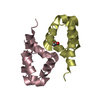
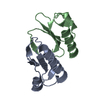
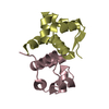
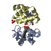
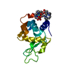
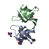
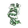
| Title | Structure of elastase inhibitor AFUEI (crystal form II) | ||||||
 Components Components | Elastase inhibitor AFUEI | ||||||
 Keywords Keywords | HYDROLASE INHIBITOR / elastase inhibitor / secreted protein | ||||||
| Function / homology |  Function and homology information Function and homology informationenzyme inhibitor activity / serine-type endopeptidase inhibitor activity / extracellular region Similarity search - Function | ||||||
| Biological species |  | ||||||
| Method |  X-RAY DIFFRACTION / SYNCHROTRON / MOLECULAR REPLACEMENT / Resolution: 1.8 Å X-RAY DIFFRACTION / SYNCHROTRON / MOLECULAR REPLACEMENT / Resolution: 1.8 Å | ||||||
 Authors Authors | Imada, K. / Sakuma, M. / Okumura, Y. / Ogawa, K. / Nikai, T. / Homma, M. | ||||||
 Citation Citation | Journal: J.Biol.Chem. / Year: 2013 Title: X-ray Structure Analysis and Characterization of AFUEI, an Elastase Inhibitor from Aspergillus fumigatus Authors: Sakuma, M. / Imada, K. / Okumura, Y. / Uchiya, K. / Yamashita, N. / Ogawa, K. / Hijikata, A. / Shirai, T. / Homma, M. / Nikai, T. | ||||||
| History |
|
- Structure visualization
| Structure viewer | Molecule: MolmilJmol/JSmol |
|---|
- Downloads & links
-Download
| PDBx/mmCIF format | 3w0e.cif.gz | 42 KB | Display | PDBx/mmCIF format |
|---|---|---|---|---|
| PDB format | pdb3w0e.ent.gz | 29.7 KB | Display | PDB format |
| PDBx/mmJSON format | 3w0e.json.gz | Tree view | PDBx/mmJSON format | |
| Others |  Other downloads Other downloads |
-Validation report
| Arichive directory | https://data.pdbj.org/pub/pdb/validation_reports/w0/3w0eftp://data.pdbj.org/pub/pdb/validation_reports/w0/3w0e | HTTPS FTP |
|---|
-Related structure data
| Related structure data |  3w0dSC S: Starting model for refinement C: citing same article ( |
|---|---|
| Similar structure data |

-Links
 PDBj
PDBj- Assembly
| Deposited unit | 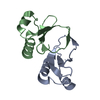
| |||||||||||||||
|---|---|---|---|---|---|---|---|---|---|---|---|---|---|---|---|---|
| 1 |
| |||||||||||||||
| Unit cell |
| |||||||||||||||
| Components on special symmetry positions |
|
-Components
| #1: Protein | Mass: 7531.449 Da / Num. of mol.: 2 / Fragment: Mature AFUEI, UNP residues 20-87 / Source method: isolated from a natural source / Source: (natural) #2: Water | ChemComp-HOH / |  Mass: 18.015 Da / Num. of mol.: 174 / Source method: isolated from a natural source / Formula: H2O Mass: 18.015 Da / Num. of mol.: 174 / Source method: isolated from a natural source / Formula: H2OHas protein modification | Y | |
|---|
-Experimental details
-Experiment
| Experiment | Method: X-RAY DIFFRACTION / Number of used crystals: 1 |
|---|
- Sample preparation
| Crystal | Density Matthews: 2.21 Å3/Da / Density % sol: 44.36 % |
|---|---|
| Crystal grow | Temperature: 293 K / Method: vapor diffusion, sitting drop / pH: 4 Details: 0.8M Sodium Sulfate, 0.1M Sodium acetate, pH 4.0, VAPOR DIFFUSION, SITTING DROP, temperature 293K |
-Data collection
| Diffraction | Mean temperature: 90 K |
|---|---|
| Diffraction source | Source: SYNCHROTRON / Site: SPring-8  / Beamline: BL41XU / Wavelength: 1 Å / Beamline: BL41XU / Wavelength: 1 Å |
| Detector | Type: RAYONIX MX225HE / Detector: CCD / Date: Jan 26, 2012 |
| Radiation | Monochromator: Double-crystal monochromator / Protocol: SINGLE WAVELENGTH / Monochromatic (M) / Laue (L): M / Scattering type: x-ray |
| Radiation wavelength | Wavelength: 1 Å / Relative weight: 1 |
| Reflection | Resolution: 1.8→32.22 Å / Num. all: 12564 / Num. obs: 12564 / % possible obs: 99.8 % / Observed criterion σ(F): 0 / Observed criterion σ(I): 0 / Redundancy: 9.1 % / Biso Wilson estimate: 22.9 Å2 / Rmerge(I) obs: 0.08 / Net I/σ(I): 17.4 |
| Reflection shell | Resolution: 1.8→1.9 Å / Redundancy: 8.9 % / Rmerge(I) obs: 0.401 / Mean I/σ(I) obs: 5.2 / Num. unique all: 1813 / % possible all: 100 |
- Processing
| Software |
| ||||||||||||||||||||||||||||||||||||||||||||||||||||||||||||
|---|---|---|---|---|---|---|---|---|---|---|---|---|---|---|---|---|---|---|---|---|---|---|---|---|---|---|---|---|---|---|---|---|---|---|---|---|---|---|---|---|---|---|---|---|---|---|---|---|---|---|---|---|---|---|---|---|---|---|---|---|---|
| Refinement | Method to determine structure: MOLECULAR REPLACEMENT Starting model: PDB ENTRY 3W0D Resolution: 1.8→32.219 Å / SU ML: 0.45 / Isotropic thermal model: isotropic / Cross valid method: THROUGHOUT / σ(F): 1.34 / Phase error: 25.56 / Stereochemistry target values: ML / Details: BULK SOLVENT MODEL USED
| ||||||||||||||||||||||||||||||||||||||||||||||||||||||||||||
| Solvent computation | Shrinkage radii: 0.72 Å / VDW probe radii: 1 Å / Solvent model: FLAT BULK SOLVENT MODEL / Bsol: 33.688 Å2 / ksol: 0.353 e/Å3 | ||||||||||||||||||||||||||||||||||||||||||||||||||||||||||||
| Displacement parameters | Biso mean: 26.8 Å2
| ||||||||||||||||||||||||||||||||||||||||||||||||||||||||||||
| Refinement step | Cycle: LAST / Resolution: 1.8→32.219 Å
| ||||||||||||||||||||||||||||||||||||||||||||||||||||||||||||
| Refine LS restraints |
| ||||||||||||||||||||||||||||||||||||||||||||||||||||||||||||
| LS refinement shell | Refine-ID: X-RAY DIFFRACTION / Total num. of bins used: 9
|
