Mass: 18.015 Da / Num. of mol.: 158 / Source method: isolated from a natural source / Formula: D2O
-
Details
Has protein modification
Y
Sequence details
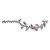
BIVALIRUDIN IS HYDROLYZED AT ARG3-PRO4 BOND DURING CRYSTALLIZATION AND BECOMES TWO CHAINS. THE ...BIVALIRUDIN IS HYDROLYZED AT ARG3-PRO4 BOND DURING CRYSTALLIZATION AND BECOMES TWO CHAINS. THE COMPLETE SEQUENCE OF BIVALIRUDIN IS (DPN)PRPGGGGNGDFEEIPEEYL.
-
Experimental details
-
Experiment
Experiment
Method
Number of used crystals
X-RAY DIFFRACTION
1
NEUTRON DIFFRACTION
1
-
Sample preparation
Crystal
ID
Density Matthews (Å3/Da)
Density % sol (%)
1
2.78
55.7
2
Crystal grow
Temperature: 298 K / Method: vapor diffusion, sitting drop / pH: 5 Details: THE CRYSTAL WAS OBTAINED BY A SITTING DROP VAPOR DIFFUSION AFTER MACROSEEDING. 2% (W/V) TO 10% (W/V) PEG4000, 100MM SODIUM ACETATE PD5.0. THE INITIAL CONCENTRATION OF THROMBIN-BIVALIRUDIN ...Details: THE CRYSTAL WAS OBTAINED BY A SITTING DROP VAPOR DIFFUSION AFTER MACROSEEDING. 2% (W/V) TO 10% (W/V) PEG4000, 100MM SODIUM ACETATE PD5.0. THE INITIAL CONCENTRATION OF THROMBIN-BIVALIRUDIN COMPLEX WAS 5MG/ML, pH 5.00, VAPOR DIFFUSION, SITTING DROP, temperature 298K
-
Data collection
Diffraction
ID
Mean temperature (K)
Crystal-ID
1
298
1
2
298
2
Diffraction source
Source
Site
Beamline
ID
Wavelength (Å)
SYNCHROTRON
Photon Factory
BL-6A
1
1
NUCLEAR REACTOR
JRR-3M
1G-C
2
2.6
Detector
Type
ID
Detector
Date
Details (eV)
ADSC QUANTUM 4r
1
CCD
Jun 13, 2009
Bendingmagnet, monochromator, mirrors
BIX-4
2
IMAGE PLATE
Mar 23, 2009
monochromator
Radiation
ID
Monochromator
Protocol
Monochromatic (M) / Laue (L)
Scattering type
Wavelength-ID
1
Si(111)
SINGLEWAVELENGTH
M
x-ray
1
2
Si(111)
M
neutron
1
Radiation wavelength
ID
Wavelength (Å)
Relative weight
1
1
1
2
2.6
1
Reflection
Entry-ID: 3VXF
Resolution (Å)
Num. all
Num. obs
% possible obs (%)
Observed criterion σ(F)
Observed criterion σ(I)
Rmerge(I) obs
Diffraction-ID
Net I/σ(I)
Biso Wilson estimate (Å2)
1.6-50
51182
51182
95.6
0
-3
0.051
1
34.5
2.8-100
8509
80
0
0.134
2
4.79
20.03
Reflection shell
Resolution (Å)
Rmerge(I) obs
Mean I/σ(I) obs
Diffraction-ID
% possible all
1.6-1.66
0.332
5.2
1
97.1
1.66-1.72
0.243
6.8
1
97.3
1.72-1.8
0.173
10.9
1
97.6
1.8-1.9
0.125
13.8
1
97.6
1.9-2.02
0.088
17.9
1
98.1
2.02-2.17
0.068
22.7
1
98.5
2.17-2.39
0.056
29.1
1
98.9
2.39-2.74
0.047
39.7
1
98.8
2.74-3.45
0.04
44.9
1
98
3.45-50
0.036
45.6
1
75.3
2.8-2.9
0.33
1.81
2
70.1
-
Processing
Software
Name
Version
Classification
PHENIX
(PHENIX.REFINE: DEV_663)
refinement
DENZO
datareduction
SCALEPACK
datascaling
Refinement
Stereochemistry target values: ML / Solvent model: FLAT BULK SOLVENT MODEL
Resolution (Å)
Refine-ID
Biso mean (Å2)
Baniso 11 (Å2)
Baniso 12 (Å2)
Baniso 13 (Å2)
Baniso 22 (Å2)
Baniso 23 (Å2)
Baniso 33 (Å2)
Rfactor Rfree
Rfactor Rwork
Rfactor obs
Num. reflection Rfree
Num. reflection all
Num. reflection obs
% reflection Rfree (%)
% reflection obs (%)
FOM work R set
SU ML
Diffraction-ID
σ(F)
Phase error
Shrinkage radii (Å)
VDW probe radii (Å)
Bsol (Å2)
ksol (e/Å3)
1.602-29.227
X-RAY DIFFRACTION
17.899
-1.0167
0
0
2.9256
-0
-1.9089
0.1842
0.1609
0.1619
1902
51136
51136
3.72
95.32
0.8582
0.16
1
1.35
16.97
1.37
1.4
53.565
0.425
2.752-44.873
NEUTRONDIFFRACTION
15.603
0.7967
-0
-0
-2.7111
0
1.9144
0.2339
0.1828
0.1855
404
8072
5
73.72
0.28
2
21.09
1.01
1.1
15.603
0.558
Refinement step
Cycle: LAST / Resolution: 1.602→29.227 Å
Protein
Nucleic acid
Ligand
Solvent
Total
Num. atoms
2357
0
14
158
2529
Refine LS restraints
Refine-ID
Type
Dev ideal
Number
NEUTRONDIFFRACTION
f_bond_d
0.016
5140
NEUTRONDIFFRACTION
f_angle_d
2.044
9130
NEUTRONDIFFRACTION
f_dihedral_angle_d
17.995
1339
NEUTRONDIFFRACTION
f_chiral_restr
0.117
350
NEUTRONDIFFRACTION
f_plane_restr
0.01
994
LS refinement shell
Resolution (Å)
Rfactor Rfree
Num. reflection Rfree
Rfactor Rwork
Num. reflection Rwork
Refine-ID
% reflection obs (%)
1.602-1.6593
0.2136
170
0.1973
4768
X-RAY DIFFRACTION
94
1.6593-1.7257
0.1875
181
0.1769
4972
X-RAY DIFFRACTION
97
1.7257-1.8042
0.2174
182
0.1676
5002
X-RAY DIFFRACTION
98
1.8042-1.8993
0.2017
182
0.163
5008
X-RAY DIFFRACTION
98
1.8993-2.0183
0.1898
183
0.1524
5010
X-RAY DIFFRACTION
98
2.0183-2.1741
0.1775
185
0.148
5077
X-RAY DIFFRACTION
98
2.1741-2.3928
0.1665
186
0.1474
5112
X-RAY DIFFRACTION
99
2.3928-2.7388
0.1625
186
0.1538
5130
X-RAY DIFFRACTION
99
2.7388-3.4498
0.1914
241
0.1613
5083
X-RAY DIFFRACTION
98
3.4498-29.2321
0.185
206
0.171
4072
X-RAY DIFFRACTION
76
2.7523-3.1505
0.2738
106
0.194
2044
NEUTRONDIFFRACTION
60
3.1505-3.9689
0.2338
141
0.1785
2705
NEUTRONDIFFRACTION
79
3.9689-44.8787
0.2171
157
0.181
2919
NEUTRONDIFFRACTION
81
+
About Yorodumi
-
News
-
Feb 9, 2022. New format data for meta-information of EMDB entries
New format data for meta-information of EMDB entries
Version 3 of the EMDB header file is now the official format.
The previous official version 1.9 will be removed from the archive.
In the structure databanks used in Yorodumi, some data are registered as the other names, "COVID-19 virus" and "2019-nCoV". Here are the details of the virus and the list of structure data.
Jan 31, 2019. EMDB accession codes are about to change! (news from PDBe EMDB page)
EMDB accession codes are about to change! (news from PDBe EMDB page)
The allocation of 4 digits for EMDB accession codes will soon come to an end. Whilst these codes will remain in use, new EMDB accession codes will include an additional digit and will expand incrementally as the available range of codes is exhausted. The current 4-digit format prefixed with “EMD-” (i.e. EMD-XXXX) will advance to a 5-digit format (i.e. EMD-XXXXX), and so on. It is currently estimated that the 4-digit codes will be depleted around Spring 2019, at which point the 5-digit format will come into force.
The EM Navigator/Yorodumi systems omit the EMD- prefix.
Related info.:Q: What is EMD? / ID/Accession-code notation in Yorodumi/EM Navigator
Yorodumi is a browser for structure data from EMDB, PDB, SASBDB, etc.
This page is also the successor to EM Navigator detail page, and also detail information page/front-end page for Omokage search.
The word "yorodu" (or yorozu) is an old Japanese word meaning "ten thousand". "mi" (miru) is to see.
Related info.:EMDB / PDB / SASBDB / Comparison of 3 databanks / Yorodumi Search / Aug 31, 2016. New EM Navigator & Yorodumi / Yorodumi Papers / Jmol/JSmol / Function and homology information / Changes in new EM Navigator and Yorodumi
 Movie
Movie Controller

 Yorodumi
 Open data
 Basic information
 Components
 Keywords
 Function and homology information
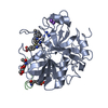
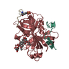
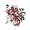
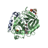
 Homo sapiens (human)
 X-RAY DIFFRACTION / NEUTRON DIFFRACTION /
  Authors
 Citation
 Structure visualization
 Downloads & links
 Other downloads

 PDBj
PDBj


 Assembly
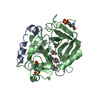
 Type: Peptide-like / Class: Thrombin inhibitor / Mass: 1779.813 Da / Num. of mol.: 1 / Fragment: C-TERMINAL / Source method: obtained synthetically
 Type: Peptide-like / Class: Thrombin inhibitor / Mass: 419.498 Da / Num. of mol.: 1 / Fragment: N-TERMINAL / Source method: obtained synthetically

 Type: D-saccharide, beta linking / Mass: 221.208 Da / Num. of mol.: 1
 Mass: 18.015 Da / Num. of mol.: 158 / Source method: isolated from a natural source / Formula: D2O
 Sample preparation
 Processing