 Movie
Movie Controller

[English] 日本語
 Yorodumi
- PDB-3vvk: An M-like Reaction State of the azide-bound purple form of pharao... -
+ Open data

- Basic information
| Entry | Database: PDB / ID: 3vvk | ||||||
|---|---|---|---|---|---|---|---|
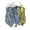
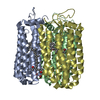
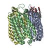
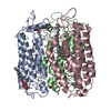
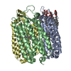
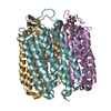
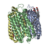
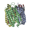
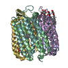
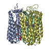
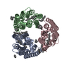
| Title | An M-like Reaction State of the azide-bound purple form of pharaonis halorhodopsin | ||||||
 Components Components | Halorhodopsin | ||||||
 Keywords Keywords | MEMBRANE PROTEIN / seven-transmembrane-retinylidene protein / chloride-bound purple form / light-driven chloride ion pump / azide-bound purple form / light-driven proton pump | ||||||
| Function / homology |  Function and homology information Function and homology informationmonoatomic ion channel activity / photoreceptor activity / phototransduction / plasma membrane Similarity search - Function | ||||||
| Biological species |  Natronomonas pharaonis (archaea) Natronomonas pharaonis (archaea) | ||||||
| Method |  X-RAY DIFFRACTION / SYNCHROTRON / MOLECULAR REPLACEMENT / Resolution: 2.3 Å X-RAY DIFFRACTION / SYNCHROTRON / MOLECULAR REPLACEMENT / Resolution: 2.3 Å | ||||||
 Authors Authors | Kouyama, T. / Nakanishi, T. | ||||||
 Citation Citation | Journal: Biophys.J. / Year: 2013 Title: Large deformation of helix F during the photoreaction cycle of Pharaonis halorhodopsin in complex with azide Authors: Nakanishi, T. / Kanada, S. / Murakami, M. / Ihara, K. / Kouyama, T. | ||||||
| History |
|
- Structure visualization
| Structure viewer | Molecule: MolmilJmol/JSmol |
|---|
- Downloads & links
-Download
| PDBx/mmCIF format | 3vvk.cif.gz | 308.4 KB | Display | PDBx/mmCIF format |
|---|---|---|---|---|
| PDB format | pdb3vvk.ent.gz | 248 KB | Display | PDB format |
| PDBx/mmJSON format | 3vvk.json.gz | Tree view | PDBx/mmJSON format | |
| Others |  Other downloads Other downloads |
-Validation report
| Summary document | 3vvk_validation.pdf.gz | 2.8 MB | Display | wwPDB validaton report |
|---|---|---|---|---|
| Full document | 3vvk_full_validation.pdf.gz | 2.9 MB | Display | |
| Data in XML | 3vvk_validation.xml.gz | 66.1 KB | Display | |
| Data in CIF | 3vvk_validation.cif.gz | 86.4 KB | Display | |
| Arichive directory | https://data.pdbj.org/pub/pdb/validation_reports/vv/3vvkftp://data.pdbj.org/pub/pdb/validation_reports/vv/3vvk | HTTPS FTP |
-Related structure data
| Related structure data |  3a7kS S: Starting model for refinement |
|---|---|
| Similar structure data |


-Links
 PDBj
PDBj


- Assembly
| Deposited unit | 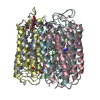
| ||||||||
|---|---|---|---|---|---|---|---|---|---|
| 1 | 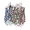
| ||||||||
| 2 | 
| ||||||||
| Unit cell |
|
-Components
-Protein / Sugars , 2 types, 10 molecules ABCDEF
| #1: Protein | Mass: 30975.096 Da / Num. of mol.: 6 / Source method: isolated from a natural source Details: STRAIN MK-1 WAS A HALORHODOPSIN-OVERPRODUCING MUTANT GENERATED FROM TYPE STRAIN D2160T. Source: (natural) Natronomonas pharaonis (archaea) / Strain: MK-1 / References: UniProt: Q3ITX1, UniProt: P15647*PLUS#3: Sugar | ChemComp-BNG /  Type: D-saccharide / Mass: 306.395 Da / Num. of mol.: 4 Type: D-saccharide / Mass: 306.395 Da / Num. of mol.: 4Source method: isolated from a genetically manipulated source Formula: C15H30O6 / Comment: detergent*YM |
|---|
-Non-polymers , 5 types, 300 molecules 


| #2: Chemical | ChemComp-RET /  Mass: 284.436 Da / Num. of mol.: 6 / Source method: obtained synthetically / Formula: C20H28O Mass: 284.436 Da / Num. of mol.: 6 / Source method: obtained synthetically / Formula: C20H28O#4: Chemical |  Mass: 741.136 Da / Num. of mol.: 2 / Source method: obtained synthetically / Formula: C50H76O4 Mass: 741.136 Da / Num. of mol.: 2 / Source method: obtained synthetically / Formula: C50H76O4#5: Chemical | ChemComp-L3P /  Mass: 885.179 Da / Num. of mol.: 4 / Source method: obtained synthetically / Formula: C46H94O11P2 Mass: 885.179 Da / Num. of mol.: 4 / Source method: obtained synthetically / Formula: C46H94O11P2#6: Chemical |  Mass: 42.020 Da / Num. of mol.: 3 / Source method: obtained synthetically / Formula: N3 Mass: 42.020 Da / Num. of mol.: 3 / Source method: obtained synthetically / Formula: N3#7: Water | ChemComp-HOH / | Mass: 18.015 Da / Num. of mol.: 285 / Source method: isolated from a natural source / Formula: H2O |
|---|
-Details
| Has protein modification | Y |
|---|
-Experimental details
-Experiment
| Experiment | Method: X-RAY DIFFRACTION / Number of used crystals: 1 |
|---|
- Sample preparation
| Crystal | Density Matthews: 1.6 Å3/Da / Density % sol: 23.17 % |
|---|---|
| Crystal grow | Temperature: 293 K / Method: vapor diffusion, sitting drop / pH: 7 Details: 5 mg/ml nonyl glucoside, 2.7M ammonium sulfate, 0.16M NaCl, 0.1M HEPES, 0.04% NaN3 , VAPOR DIFFUSION, SITTING DROP, temperature 293K |
-Data collection
| Diffraction | Mean temperature: 100 K | |||||||||||||||||||||
|---|---|---|---|---|---|---|---|---|---|---|---|---|---|---|---|---|---|---|---|---|---|---|
| Diffraction source | Source: SYNCHROTRON / Site: SPring-8  / Beamline: BL38B1 / Wavelength: 1 Å / Beamline: BL38B1 / Wavelength: 1 Å | |||||||||||||||||||||
| Detector | Type: ADSC QUANTUM 315 / Detector: CCD / Date: Feb 17, 2011 / Details: mirrors | |||||||||||||||||||||
| Radiation | Monochromator: Si (111) double crystal monochromator / Protocol: SINGLE WAVELENGTH / Monochromatic (M) / Laue (L): M / Scattering type: x-ray | |||||||||||||||||||||
| Radiation wavelength | Wavelength: 1 Å / Relative weight: 1 | |||||||||||||||||||||
| Reflection | Resolution: 2.3→79 Å / Num. all: 52756 / Num. obs: 48183 / % possible obs: 92.5 % / Observed criterion σ(F): 3 / Observed criterion σ(I): 3 / Redundancy: 3.6 % / Biso Wilson estimate: 28.2 Å2 / Rmerge(I) obs: 0.097 / Rsym value: 0.097 / Net I/σ(I): 9.6 | |||||||||||||||||||||
| Reflection shell | Diffraction-ID: 1
|
- Processing
| Software |
| |||||||||||||||||||||||||
|---|---|---|---|---|---|---|---|---|---|---|---|---|---|---|---|---|---|---|---|---|---|---|---|---|---|---|
| Refinement | Method to determine structure: MOLECULAR REPLACEMENT Starting model: 3a7k Resolution: 2.3→15 Å / Occupancy max: 1 / Occupancy min: 0.36 / Isotropic thermal model: Overall / Cross valid method: THROUGHOUT / σ(F): 0 / Stereochemistry target values: Engh & Huber Details: THE POLYPEPTIDE CHAINS A, B AND C REPRESENT THE REACTION STATES TRAPED IN THE THREE SUBUNITS WITHIN THE ASYMMETRIC UNIT WHEN THE C2 CRYSTAL OF THE HALORHODOPSIN-AZIDE COMPLEX WAS FLASH- ...Details: THE POLYPEPTIDE CHAINS A, B AND C REPRESENT THE REACTION STATES TRAPED IN THE THREE SUBUNITS WITHIN THE ASYMMETRIC UNIT WHEN THE C2 CRYSTAL OF THE HALORHODOPSIN-AZIDE COMPLEX WAS FLASH-COOLED UNDER THE ILLUMINATION WITH ORANGE LIGHT, WHILE THE CHAINS D, E AND F REPRESENT THE UNPHOTOLYSED STATES.
| |||||||||||||||||||||||||
| Solvent computation | Bsol: 75.1402 Å2 | |||||||||||||||||||||||||
| Displacement parameters | Biso max: 77.46 Å2 / Biso mean: 26.524 Å2 / Biso min: 0 Å2
| |||||||||||||||||||||||||
| Refine analyze |
| |||||||||||||||||||||||||
| Refinement step | Cycle: LAST / Resolution: 2.3→15 Å
| |||||||||||||||||||||||||
| Refine LS restraints |
| |||||||||||||||||||||||||
| LS refinement shell | Resolution: 2.3→2.38 Å
| |||||||||||||||||||||||||
| Xplor file |
|
