 Movie
Movie Controller


+ Open data

- Basic information
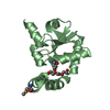
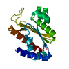
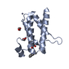
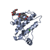
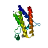
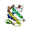
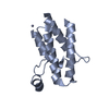
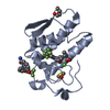
| Entry | Database: PDB / ID: 3vts | ||||||
|---|---|---|---|---|---|---|---|
| Title | Crystal structure of a three finger toxin from snake venom | ||||||
 Components Components | Cytotoxin 1 | ||||||
 Keywords Keywords | TOXIN / Three finger toxin / Venom toxin | ||||||
| Function / homology |  Function and homology information Function and homology informationregulation of blood pressure / toxin activity / killing of cells of another organism / extracellular region Similarity search - Function | ||||||
| Biological species |  Hemachatus haemachatus (ringhals) Hemachatus haemachatus (ringhals) | ||||||
| Method |  X-RAY DIFFRACTION / MOLECULAR REPLACEMENT / Resolution: 2.426 Å X-RAY DIFFRACTION / MOLECULAR REPLACEMENT / Resolution: 2.426 Å | ||||||
 Authors Authors | Jobichen, C. / Sivaraman, J. | ||||||
 Citation Citation | Journal: Plos One / Year: 2012 Title: Identification and structural characterization of a new three-finger toxin hemachatoxin from Hemachatus haemachatus venom. Authors: Girish, V.M. / Kumar, S. / Joseph, L. / Jobichen, C. / Kini, R.M. / Sivaraman, J. | ||||||
| History |
|
- Structure visualization
| Structure viewer | Molecule: MolmilJmol/JSmol |
|---|
- Downloads & links
-Download
| PDBx/mmCIF format | 3vts.cif.gz | 58.9 KB | Display | PDBx/mmCIF format |
|---|---|---|---|---|
| PDB format | pdb3vts.ent.gz | 44 KB | Display | PDB format |
| PDBx/mmJSON format | 3vts.json.gz | Tree view | PDBx/mmJSON format | |
| Others |  Other downloads Other downloads |
-Validation report
| Arichive directory | https://data.pdbj.org/pub/pdb/validation_reports/vt/3vtsftp://data.pdbj.org/pub/pdb/validation_reports/vt/3vts | HTTPS FTP |
|---|
-Related structure data
| Related structure data |  1tgxS S: Starting model for refinement |
|---|---|
| Similar structure data |


-Links
 PDBj
PDBj

- Assembly
| Deposited unit | 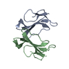
| ||||||||||||||||||
|---|---|---|---|---|---|---|---|---|---|---|---|---|---|---|---|---|---|---|---|
| 1 |
| ||||||||||||||||||
| Unit cell |
| ||||||||||||||||||
| Noncrystallographic symmetry (NCS) | NCS domain:
NCS domain segments:
|
-Components
| #1: Protein | Mass: 6858.533 Da / Num. of mol.: 2 / Source method: isolated from a natural source / Source: (natural) Hemachatus haemachatus (ringhals) / References: UniProt: B3EWH9*PLUS#2: Water | ChemComp-HOH / |  Mass: 18.015 Da / Num. of mol.: 49 / Source method: isolated from a natural source / Formula: H2O Mass: 18.015 Da / Num. of mol.: 49 / Source method: isolated from a natural source / Formula: H2OHas protein modification | Y | Sequence details | THE AUTHOR DEPOSITED SEQUENCE OF THIS PROTEIN AS THE CODE B3EWH9 BUT IT DOES NOT APPEAR IN TE ...THE AUTHOR DEPOSITED SEQUENCE OF THIS PROTEIN AS THE CODE B3EWH9 BUT IT DOES NOT APPEAR IN TE SEQUENCE DATABASE YET. | |
|---|
-Experimental details
-Experiment
| Experiment | Method: X-RAY DIFFRACTION / Number of used crystals: 1 |
|---|
- Sample preparation
| Crystal | Density Matthews: 2.62 Å3/Da / Density % sol: 53.09 % |
|---|---|
| Crystal grow | Temperature: 298 K / Method: vapor diffusion, hanging drop / pH: 4.5 Details: 150mM ammonium acetate, 100mM sodium acetate, 25% polyethylene glycol 4000 , pH 4.5, VAPOR DIFFUSION, HANGING DROP, temperature 298K |
-Data collection
| Diffraction | Mean temperature: 100 K |
|---|---|
| Diffraction source | Source: ROTATING ANODE / Type: RIGAKU / Wavelength: 1.5418 Å |
| Detector | Type: RIGAKU RAXIS IV++ / Detector: IMAGE PLATE / Date: Oct 31, 2008 / Details: mirrors |
| Radiation | Monochromator: GRAPHITE / Protocol: SINGLE WAVELENGTH / Monochromatic (M) / Laue (L): M / Scattering type: x-ray |
| Radiation wavelength | Wavelength: 1.5418 Å / Relative weight: 1 |
| Reflection | Resolution: 2.426→30.112 Å / Num. all: 5639 / Num. obs: 5614 / % possible obs: 96.2 % / Observed criterion σ(F): 2 / Observed criterion σ(I): 2 / Redundancy: 3.9 % / Rmerge(I) obs: 0.064 / Net I/σ(I): 20.6 |
- Processing
| Software |
| ||||||||||||||||||||||||||||||||||||||||
|---|---|---|---|---|---|---|---|---|---|---|---|---|---|---|---|---|---|---|---|---|---|---|---|---|---|---|---|---|---|---|---|---|---|---|---|---|---|---|---|---|---|
| Refinement | Method to determine structure: MOLECULAR REPLACEMENT Starting model: 1TGX Resolution: 2.426→30.112 Å / SU ML: 0.41 / σ(F): 1.35 / Phase error: 29.36 / Stereochemistry target values: ML
| ||||||||||||||||||||||||||||||||||||||||
| Solvent computation | Shrinkage radii: 0.98 Å / VDW probe radii: 1.2 Å / Solvent model: FLAT BULK SOLVENT MODEL / Bsol: 19.328 Å2 / ksol: 0.29 e/Å3 | ||||||||||||||||||||||||||||||||||||||||
| Displacement parameters |
| ||||||||||||||||||||||||||||||||||||||||
| Refinement step | Cycle: LAST / Resolution: 2.426→30.112 Å
| ||||||||||||||||||||||||||||||||||||||||
| Refine LS restraints |
| ||||||||||||||||||||||||||||||||||||||||
| Refine LS restraints NCS |
| ||||||||||||||||||||||||||||||||||||||||
| LS refinement shell | Refine-ID: X-RAY DIFFRACTION / Total num. of bins used: 4
| ||||||||||||||||||||||||||||||||||||||||
| Refinement TLS params. | Method: refined / Origin x: 61.4575 Å / Origin y: 56.864 Å / Origin z: 9.017 Å
| ||||||||||||||||||||||||||||||||||||||||
| Refinement TLS group | Selection details: ALL |
