 Movie
Movie Controller


+ Open data

- Basic information
| Entry | Database: PDB / ID: 3vsv | |||||||||
|---|---|---|---|---|---|---|---|---|---|---|
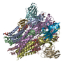
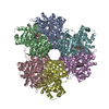
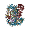
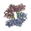
| Title | The complex structure of XylC with xylose | |||||||||
 Components Components | Xylosidase | |||||||||
 Keywords Keywords | HYDROLASE / glycoside hydrolase / beta-xylosidase / product inhibition | |||||||||
| Function / homology |  Function and homology information Function and homology informationcarbon-oxygen lyase activity, acting on polysaccharides / extracellular region Similarity search - Function | |||||||||
| Biological species |  Thermoanaerobacterium (bacteria) Thermoanaerobacterium (bacteria) | |||||||||
| Method |  X-RAY DIFFRACTION / SYNCHROTRON / MOLECULAR REPLACEMENT / Resolution: 1.48 Å X-RAY DIFFRACTION / SYNCHROTRON / MOLECULAR REPLACEMENT / Resolution: 1.48 Å | |||||||||
 Authors Authors | Huang, C.H. / Sun, Y. / Ko, T.P. / Ma, Y. / Chen, C.C. / Zheng, Y. / Chan, H.C. / Pang, X. / Wiegel, J. / Shao, W. / Guo, R.T. | |||||||||
 Citation Citation | Journal: Biochem.J. / Year: 2012 Title: The substrate/product-binding modes of a novel GH120 beta-xylosidase (XylC) from Thermoanaerobacterium saccharolyticum JW/SL-YS485 Authors: Huang, C.H. / Sun, Y. / Ko, T.P. / Chen, C.C. / Zheng, Y. / Chan, H.C. / Pang, X. / Wiegel, J. / Shao, W. / Guo, R.T. | |||||||||
| History |
|
- Structure visualization
| Structure viewer | Molecule: MolmilJmol/JSmol |
|---|
- Downloads & links
-Download
| PDBx/mmCIF format | 3vsv.cif.gz | 597.1 KB | Display | PDBx/mmCIF format |
|---|---|---|---|---|
| PDB format | pdb3vsv.ent.gz | 486.8 KB | Display | PDB format |
| PDBx/mmJSON format | 3vsv.json.gz | Tree view | PDBx/mmJSON format | |
| Others |  Other downloads Other downloads |
-Validation report
| Arichive directory | https://data.pdbj.org/pub/pdb/validation_reports/vs/3vsvftp://data.pdbj.org/pub/pdb/validation_reports/vs/3vsv | HTTPS FTP |
|---|
-Related structure data
| Related structure data |  3vstSC  3vsuC S: Starting model for refinement C: citing same article ( |
|---|---|
| Similar structure data |

-Links
 PDBj
PDBj- Assembly
| Deposited unit | 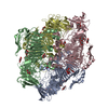
| ||||||||
|---|---|---|---|---|---|---|---|---|---|
| 1 |
| ||||||||
| Unit cell |
|
-Components
| #1: Protein | Mass: 72845.398 Da / Num. of mol.: 4 Source method: isolated from a genetically manipulated source Source: (gene. exp.) Thermoanaerobacterium (bacteria) / Strain: JW/SL YS485 / Plasmid: pHsh / Production host: #2: Sugar | ChemComp-XYP /   Type: D-saccharide, beta linking / Mass: 150.130 Da / Num. of mol.: 26 Type: D-saccharide, beta linking / Mass: 150.130 Da / Num. of mol.: 26Source method: isolated from a genetically manipulated source Formula: C5H10O5 #3: Sugar | ChemComp-XYS /   Type: D-saccharide, alpha linking / Mass: 150.130 Da / Num. of mol.: 9 Type: D-saccharide, alpha linking / Mass: 150.130 Da / Num. of mol.: 9Source method: isolated from a genetically manipulated source Formula: C5H10O5 #4: Water | ChemComp-HOH / |  Mass: 18.015 Da / Num. of mol.: 3613 / Source method: isolated from a natural source / Formula: H2O Mass: 18.015 Da / Num. of mol.: 3613 / Source method: isolated from a natural source / Formula: H2O |
|---|
-Experimental details
-Experiment
| Experiment | Method: X-RAY DIFFRACTION |
|---|
- Sample preparation
| Crystal | Density Matthews: 3.04 Å3/Da / Density % sol: 59.54 % |
|---|---|
| Crystal grow | Temperature: 295 K / Method: vapor diffusion, sitting drop / pH: 5.6 Details: 0.2M sodium citrate, 15-17%(w/v) polyethylene glycol 3350, pH 5.6, VAPOR DIFFUSION, SITTING DROP, temperature 295K |
-Data collection
| Diffraction | Mean temperature: 100 K |
|---|---|
| Diffraction source | Source: SYNCHROTRON / Site: NSRRC  / Beamline: BL13B1 / Wavelength: 1 Å / Beamline: BL13B1 / Wavelength: 1 Å |
| Detector | Date: Mar 10, 2011 |
| Radiation | Protocol: SINGLE WAVELENGTH / Monochromatic (M) / Laue (L): M / Scattering type: x-ray |
| Radiation wavelength | Wavelength: 1 Å / Relative weight: 1 |
| Reflection | Resolution: 1.48→25 Å / Num. obs: 574432 / % possible obs: 100 % / Redundancy: 3.7 % / Rmerge(I) obs: 0.071 / Net I/σ(I): 20.9 |
| Reflection shell | Resolution: 1.48→1.53 Å / Redundancy: 3.5 % / Rmerge(I) obs: 0.496 / Mean I/σ(I) obs: 3.3 / Num. unique all: 57397 / % possible all: 100 |
- Processing
| Software |
| ||||||||||||||||||||
|---|---|---|---|---|---|---|---|---|---|---|---|---|---|---|---|---|---|---|---|---|---|
| Refinement | Method to determine structure: MOLECULAR REPLACEMENT Starting model: 3VST Resolution: 1.48→25 Å / σ(F): 2 / Stereochemistry target values: Engh & Huber
| ||||||||||||||||||||
| Refinement step | Cycle: LAST / Resolution: 1.48→25 Å
| ||||||||||||||||||||
| LS refinement shell | Resolution: 1.48→1.53 Å
|
