 Movie
Movie Controller

[English] 日本語
 Yorodumi
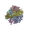
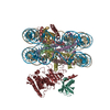
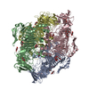
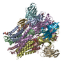
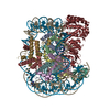
- PDB-4r8p: Crystal structure of the Ring1B/Bmi1/UbcH5c PRC1 ubiquitylation m... -
+ Open data

- Basic information
| Entry | Database: PDB / ID: 4r8p | ||||||
|---|---|---|---|---|---|---|---|
| Title | Crystal structure of the Ring1B/Bmi1/UbcH5c PRC1 ubiquitylation module bound to the nucleosome core particle | ||||||
 Components Components |
| ||||||
 Keywords Keywords | Structural Protein/DNA / RING domain / Arginine anchor / Ubiquitin ligase / Histone modification enzyme / Structural Protein-DNA complex | ||||||
| Function / homology |  Function and homology information Function and homology informationregulation of adaxial/abaxial pattern formation / histone H2AK119 ubiquitin ligase activity / segment specification / PRC1 complex / RING-like zinc finger domain binding / regulation of kidney development / rostrocaudal neural tube patterning / somatic stem cell division / embryonic skeletal system morphogenesis / ubiquitin-protein transferase activator activity ...regulation of adaxial/abaxial pattern formation / histone H2AK119 ubiquitin ligase activity / segment specification / PRC1 complex / RING-like zinc finger domain binding / regulation of kidney development / rostrocaudal neural tube patterning / somatic stem cell division / embryonic skeletal system morphogenesis / ubiquitin-protein transferase activator activity / sex chromatin / positive regulation of immature T cell proliferation in thymus / Signaling by BMP / PcG protein complex / (E3-independent) E2 ubiquitin-conjugating enzyme / gastrulation with mouth forming second / SUMOylation of DNA methylation proteins / SUMOylation of RNA binding proteins / protein K11-linked ubiquitination / protein K6-linked ubiquitination / anterior/posterior axis specification / positive regulation of ubiquitin-protein transferase activity / : / E2 ubiquitin-conjugating enzyme / DNA methylation-dependent constitutive heterochromatin formation / negative regulation of gene expression, epigenetic / germ cell development / Transcriptional Regulation by E2F6 / humoral immune response / ubiquitin conjugating enzyme activity / RUNX1 interacts with co-factors whose precise effect on RUNX1 targets is not known / MLL1 complex / negative regulation of BMP signaling pathway / hemopoiesis / negative regulation of apoptotic signaling pathway / : / cellular response to dexamethasone stimulus / cellular response to interleukin-1 / protein monoubiquitination / ubiquitin ligase complex / SUMOylation of DNA damage response and repair proteins / protein autoubiquitination / positive regulation of B cell proliferation / heterochromatin / protein K48-linked ubiquitination / SUMOylation of transcription cofactors / epigenetic regulation of gene expression / SUMOylation of chromatin organization proteins / protein modification process / Regulation of PTEN gene transcription / TICAM1, RIP1-mediated IKK complex recruitment / IKK complex recruitment mediated by RIP1 / PINK1-PRKN Mediated Mitophagy / apoptotic signaling pathway / Negative regulators of DDX58/IFIH1 signaling / Peroxisomal protein import / promoter-specific chromatin binding / Downregulation of SMAD2/3:SMAD4 transcriptional activity / brain development / euchromatin / Regulation of TNFR1 signaling / RING-type E3 ubiquitin transferase / Inactivation of CSF3 (G-CSF) signaling / positive regulation of fibroblast proliferation / Oxygen-dependent proline hydroxylation of Hypoxia-inducible Factor Alpha / protein polyubiquitination / ubiquitin-protein transferase activity / structural constituent of chromatin / ubiquitin protein ligase activity / nucleosome / mitotic cell cycle / nucleosome assembly / Antigen processing: Ubiquitination & Proteasome degradation / E3 ubiquitin ligases ubiquitinate target proteins / heterochromatin formation / in utero embryonic development / regulation of gene expression / Neddylation / gene expression / Oxidative Stress Induced Senescence / ubiquitin-dependent protein catabolic process / proteasome-mediated ubiquitin-dependent protein catabolic process / endosome membrane / nuclear body / protein ubiquitination / chromatin remodeling / protein heterodimerization activity / DNA repair / chromatin binding / apoptotic process / negative regulation of transcription by RNA polymerase II / DNA binding / extracellular exosome / zinc ion binding / nucleoplasm / ATP binding / nucleus / plasma membrane / cytosol / cytoplasm Similarity search - Function | ||||||
| Biological species | Synthetic DNA (others)  Homo sapiens (human) Homo sapiens (human) | ||||||
| Method |  X-RAY DIFFRACTION / SYNCHROTRON / MOLECULAR REPLACEMENT / molecular replacement / Resolution: 3.2846 Å X-RAY DIFFRACTION / SYNCHROTRON / MOLECULAR REPLACEMENT / molecular replacement / Resolution: 3.2846 Å | ||||||
 Authors Authors | McGinty, R.K. / Henrici, R.C. / Tan, S. | ||||||
 Citation Citation | Journal: Nature / Year: 2014 Title: Crystal structure of the PRC1 ubiquitylation module bound to the nucleosome. Authors: McGinty, R.K. / Henrici, R.C. / Tan, S. | ||||||
| History |
|
- Structure visualization
| Structure viewer | Molecule: MolmilJmol/JSmol |
|---|
- Downloads & links
-Download
| PDBx/mmCIF format | 4r8p.cif.gz | 905.5 KB | Display | PDBx/mmCIF format |
|---|---|---|---|---|
| PDB format | pdb4r8p.ent.gz | 737.8 KB | Display | PDB format |
| PDBx/mmJSON format | 4r8p.json.gz | Tree view | PDBx/mmJSON format | |
| Others |  Other downloads Other downloads |
-Validation report
| Arichive directory | https://data.pdbj.org/pub/pdb/validation_reports/r8/4r8pftp://data.pdbj.org/pub/pdb/validation_reports/r8/4r8p | HTTPS FTP |
|---|
-Related structure data


-Links
 PDBj
PDBj


- Assembly
| Deposited unit | 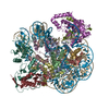
| ||||||||
|---|---|---|---|---|---|---|---|---|---|
| 1 |
| ||||||||
| Unit cell |
|
-Components
-Protein , 6 types, 12 molecules AEBFCGDHKMLN
| #1: Protein | Mass: 15303.930 Da / Num. of mol.: 2 Source method: isolated from a genetically manipulated source Source: (gene. exp.)  #2: Protein | Mass: 11263.231 Da / Num. of mol.: 2 Source method: isolated from a genetically manipulated source Source: (gene. exp.) #3: Protein | Mass: 13978.241 Da / Num. of mol.: 2 Source method: isolated from a genetically manipulated source Source: (gene. exp.) #4: Protein | Mass: 13524.752 Da / Num. of mol.: 2 Source method: isolated from a genetically manipulated source Source: (gene. exp.) #7: Protein | Mass: 12595.749 Da / Num. of mol.: 2 / Fragment: Residues 2-109 Source method: isolated from a genetically manipulated source Source: (gene. exp.) Homo sapiens (human) / Gene: BMI1, PCGF4, RNF51 / Plasmid: pST44 / Production host: #8: Protein | Mass: 30118.637 Da / Num. of mol.: 2 Fragment: Residues 2-116 of Ring1B and Residues 2-148 of UbcH5c Source method: isolated from a genetically manipulated source Details: Ring1B(2-116) fragment linked to UBE2D3/UbcH5c (2-148) through a linker encodin g the GSGSRS sequence Source: (gene. exp.) Homo sapiens (human)Gene: BAP1, DING, HIPI3, RING1B, Ring1B-UbcH5c fusion, RNF2, UBC5C, UBCH5C, UBE2D3 Plasmid: pST44 / Production host: References: UniProt: Q99496, UniProt: P61077, Ligases; Forming carbon-nitrogen bonds; Acid-amino-acid ligases (peptide synthases), ubiquitin-protein ligase |
|---|
-DNA chain , 2 types, 2 molecules IJ
| #5: DNA chain | Mass: 45138.770 Da / Num. of mol.: 1 / Fragment: Widom 601 147-mer (+ strand) Source method: isolated from a genetically manipulated source Source: (gene. exp.) Synthetic DNA (others) / Gene: Widom 601 nucleosome positioning sequence / Plasmid: pST55 / Production host: |
|---|---|
| #6: DNA chain | Mass: 45610.043 Da / Num. of mol.: 1 / Fragment: Widom 601 147-mer (- strand) Source method: isolated from a genetically manipulated source Source: (gene. exp.) Synthetic DNA (others) / Gene: Widom 601 nucleosome positioning sequence / Plasmid: pST55 / Production host: |
-Non-polymers , 1 types, 8 molecules 
| #9: Chemical | ChemComp-ZN /  Mass: 65.409 Da / Num. of mol.: 8 / Source method: obtained synthetically / Formula: Zn Mass: 65.409 Da / Num. of mol.: 8 / Source method: obtained synthetically / Formula: Zn |
|---|
-Experimental details
-Experiment
| Experiment | Method: X-RAY DIFFRACTION / Number of used crystals: 1 |
|---|
- Sample preparation
| Crystal | Density Matthews: 3.12 Å3/Da / Density % sol: 60.53 % |
|---|---|
| Crystal grow | Temperature: 294 K / Method: modified micro batch under oil / pH: 7.5 Details: 25 mM HEPES pH 7.5, 80 mM NH4NO3, 3 % PEG2000-MME, Modified micro batch under oil, temperature 294K |
-Data collection
| Diffraction | Mean temperature: 100 K | ||||||||||||||||||||||||||||||||||||||||||||||||||||||||||||||||||||||||||||||||||||||||
|---|---|---|---|---|---|---|---|---|---|---|---|---|---|---|---|---|---|---|---|---|---|---|---|---|---|---|---|---|---|---|---|---|---|---|---|---|---|---|---|---|---|---|---|---|---|---|---|---|---|---|---|---|---|---|---|---|---|---|---|---|---|---|---|---|---|---|---|---|---|---|---|---|---|---|---|---|---|---|---|---|---|---|---|---|---|---|---|---|---|
| Diffraction source | Source: SYNCHROTRON / Site: APS  / Beamline: 24-ID-E / Wavelength: 0.97918 Å / Beamline: 24-ID-E / Wavelength: 0.97918 Å | ||||||||||||||||||||||||||||||||||||||||||||||||||||||||||||||||||||||||||||||||||||||||
| Detector | Type: ADSC QUANTUM 315 / Detector: CCD / Date: Jul 29, 2013 | ||||||||||||||||||||||||||||||||||||||||||||||||||||||||||||||||||||||||||||||||||||||||
| Radiation | Monochromator: Si(220) / Protocol: SINGLE WAVELENGTH / Monochromatic (M) / Laue (L): M / Scattering type: x-ray | ||||||||||||||||||||||||||||||||||||||||||||||||||||||||||||||||||||||||||||||||||||||||
| Radiation wavelength | Wavelength: 0.97918 Å / Relative weight: 1 | ||||||||||||||||||||||||||||||||||||||||||||||||||||||||||||||||||||||||||||||||||||||||
| Reflection | Resolution: 3.2846→45 Å / Num. all: 54219 / Num. obs: 54142 / % possible obs: 99.4 % / Observed criterion σ(F): 0 / Observed criterion σ(I): -4 / Redundancy: 3.9 % / Biso Wilson estimate: 105.01 Å2 / Rsym value: 0.063 / Net I/σ(I): 16.5 | ||||||||||||||||||||||||||||||||||||||||||||||||||||||||||||||||||||||||||||||||||||||||
| Reflection shell | Diffraction-ID: 1
|
-Phasing
| Phasing | Method: molecular replacement | |||||||||
|---|---|---|---|---|---|---|---|---|---|---|
| Phasing MR | Model details: Phaser MODE: MR_AUTO
|
- Processing
| Software |
| ||||||||||||||||||||||||||||||||||||||||||||||||||||||||||||||||||||||||||||||||||||||||||||||||||||||||||||||||||||||||||||||||||||||||||||||||||||||||||||||||||||||||
|---|---|---|---|---|---|---|---|---|---|---|---|---|---|---|---|---|---|---|---|---|---|---|---|---|---|---|---|---|---|---|---|---|---|---|---|---|---|---|---|---|---|---|---|---|---|---|---|---|---|---|---|---|---|---|---|---|---|---|---|---|---|---|---|---|---|---|---|---|---|---|---|---|---|---|---|---|---|---|---|---|---|---|---|---|---|---|---|---|---|---|---|---|---|---|---|---|---|---|---|---|---|---|---|---|---|---|---|---|---|---|---|---|---|---|---|---|---|---|---|---|---|---|---|---|---|---|---|---|---|---|---|---|---|---|---|---|---|---|---|---|---|---|---|---|---|---|---|---|---|---|---|---|---|---|---|---|---|---|---|---|---|---|---|---|---|---|---|---|---|
| Refinement | Method to determine structure: MOLECULAR REPLACEMENT Starting model: polyalanine models of PDB entries 3RPG and 3LZ0 Resolution: 3.2846→45 Å / SU ML: 0.43 / Isotropic thermal model: isotropic / Cross valid method: THROUGHOUT / σ(F): 1.34 / Phase error: 25.77 / Stereochemistry target values: ML
| ||||||||||||||||||||||||||||||||||||||||||||||||||||||||||||||||||||||||||||||||||||||||||||||||||||||||||||||||||||||||||||||||||||||||||||||||||||||||||||||||||||||||
| Solvent computation | Shrinkage radii: 0.9 Å / VDW probe radii: 1.11 Å / Solvent model: FLAT BULK SOLVENT MODEL | ||||||||||||||||||||||||||||||||||||||||||||||||||||||||||||||||||||||||||||||||||||||||||||||||||||||||||||||||||||||||||||||||||||||||||||||||||||||||||||||||||||||||
| Displacement parameters | Biso max: 368.24 Å2 / Biso mean: 137.4911 Å2 / Biso min: 44.46 Å2 | ||||||||||||||||||||||||||||||||||||||||||||||||||||||||||||||||||||||||||||||||||||||||||||||||||||||||||||||||||||||||||||||||||||||||||||||||||||||||||||||||||||||||
| Refinement step | Cycle: LAST / Resolution: 3.2846→45 Å
| ||||||||||||||||||||||||||||||||||||||||||||||||||||||||||||||||||||||||||||||||||||||||||||||||||||||||||||||||||||||||||||||||||||||||||||||||||||||||||||||||||||||||
| Refine LS restraints |
| ||||||||||||||||||||||||||||||||||||||||||||||||||||||||||||||||||||||||||||||||||||||||||||||||||||||||||||||||||||||||||||||||||||||||||||||||||||||||||||||||||||||||
| LS refinement shell | Refine-ID: X-RAY DIFFRACTION / Total num. of bins used: 20
| ||||||||||||||||||||||||||||||||||||||||||||||||||||||||||||||||||||||||||||||||||||||||||||||||||||||||||||||||||||||||||||||||||||||||||||||||||||||||||||||||||||||||
| Refinement TLS params. | Method: refined / Origin x: 29.28 Å / Origin y: 30.3205 Å / Origin z: -47.343 Å
| ||||||||||||||||||||||||||||||||||||||||||||||||||||||||||||||||||||||||||||||||||||||||||||||||||||||||||||||||||||||||||||||||||||||||||||||||||||||||||||||||||||||||
| Refinement TLS group |
|
