 Movie
Movie Controller

[English] 日本語
 Yorodumi
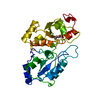
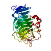
- PDB-3vmv: Crystal structure of pectate lyase Bsp165PelA from Bacillus sp. N165 -
+ Open data

- Basic information
| Entry | Database: PDB / ID: 3vmv | ||||||
|---|---|---|---|---|---|---|---|
| Title | Crystal structure of pectate lyase Bsp165PelA from Bacillus sp. N165 | ||||||
 Components Components | Pectate lyase | ||||||
 Keywords Keywords | LYASE / Polysaccharide Lyase Family 1 / beta-helix / pectolytic / polygalacturonate | ||||||
| Function / homology |  Function and homology information Function and homology informationpectate lyase activity / polysaccharide catabolic process / extracellular region Similarity search - Function | ||||||
| Biological species |  | ||||||
| Method |  X-RAY DIFFRACTION / SYNCHROTRON / MOLECULAR REPLACEMENT / Resolution: 1.54 Å X-RAY DIFFRACTION / SYNCHROTRON / MOLECULAR REPLACEMENT / Resolution: 1.54 Å | ||||||
 Authors Authors | Zheng, Y. / Huang, C.H. / Liu, W. / Ko, T.P. / Xue, Y. / Zhou, C. / Zhang, G. / Guo, R.T. / Ma, Y. | ||||||
 Citation Citation | Journal: Biochem.Biophys.Res.Commun. / Year: 2012 Title: Crystal structure and substrate-binding mode of a novel pectate lyase from alkaliphilic Bacillus sp. N16-5. Authors: Zheng, Y. / Huang, C.H. / Liu, W. / Ko, T.P. / Xue, Y. / Zhou, C. / Guo, R.T. / Ma, Y. | ||||||
| History |
|
- Structure visualization
| Structure viewer | Molecule: MolmilJmol/JSmol |
|---|
- Downloads & links
-Download
| PDBx/mmCIF format | 3vmv.cif.gz | 150.4 KB | Display | PDBx/mmCIF format |
|---|---|---|---|---|
| PDB format | pdb3vmv.ent.gz | 118.5 KB | Display | PDB format |
| PDBx/mmJSON format | 3vmv.json.gz | Tree view | PDBx/mmJSON format | |
| Others |  Other downloads Other downloads |
-Validation report
| Arichive directory | https://data.pdbj.org/pub/pdb/validation_reports/vm/3vmvftp://data.pdbj.org/pub/pdb/validation_reports/vm/3vmv | HTTPS FTP |
|---|
-Related structure data
| Related structure data | 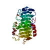 3vmwC 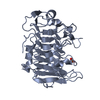 2qxzS C: citing same article ( S: Starting model for refinement |
|---|---|
| Similar structure data |


-Links
 PDBj
PDBj- Assembly
| Deposited unit | 
| ||||||||||||
|---|---|---|---|---|---|---|---|---|---|---|---|---|---|
| 1 |
| ||||||||||||
| Unit cell |
| ||||||||||||
| Components on special symmetry positions |
|
-Components
| #1: Protein | Mass: 35979.137 Da / Num. of mol.: 1 / Fragment: catalytic domain (UNP RESIDUES 37-362) Source method: isolated from a genetically manipulated source Source: (gene. exp.) | ||
|---|---|---|---|
| #2: Chemical |   Mass: 96.063 Da / Num. of mol.: 2 / Source method: obtained synthetically / Formula: SO4 Mass: 96.063 Da / Num. of mol.: 2 / Source method: obtained synthetically / Formula: SO4#3: Water | ChemComp-HOH / |  Mass: 18.015 Da / Num. of mol.: 349 / Source method: isolated from a natural source / Formula: H2O Mass: 18.015 Da / Num. of mol.: 349 / Source method: isolated from a natural source / Formula: H2O |
-Experimental details
-Experiment
| Experiment | Method: X-RAY DIFFRACTION / Number of used crystals: 1 |
|---|
- Sample preparation
| Crystal | Density Matthews: 2.4 Å3/Da / Density % sol: 48.85 % |
|---|---|
| Crystal grow | Temperature: 293 K / Method: vapor diffusion, hanging drop / pH: 8 Details: 0.18M Li2SO4, 0.085M Tris-HCl, pH 8.5, 24% PEG 4000, 15% v/v Glycerol anhydrous, VAPOR DIFFUSION, HANGING DROP, temperature 293K |
-Data collection
| Diffraction | Mean temperature: 100 K |
|---|---|
| Diffraction source | Source: SYNCHROTRON / Site: NSRRC  / Beamline: BL13C1 / Wavelength: 1 Å / Beamline: BL13C1 / Wavelength: 1 Å |
| Detector | Type: ADSC QUANTUM 315r / Detector: CCD / Date: Nov 20, 2010 |
| Radiation | Protocol: SINGLE WAVELENGTH / Monochromatic (M) / Laue (L): M / Scattering type: x-ray |
| Radiation wavelength | Wavelength: 1 Å / Relative weight: 1 |
| Reflection | Resolution: 1.54→25 Å / Num. obs: 51969 / % possible obs: 99.3 % / Observed criterion σ(I): 0 / Redundancy: 7.8 % / Rmerge(I) obs: 0.059 |
| Reflection shell | Resolution: 1.54→1.6 Å / Redundancy: 7.5 % / Rmerge(I) obs: 0.301 / Num. unique all: 38662 / % possible all: 99.8 |
- Processing
| Software |
| ||||||||||||||||||||||||||||||||||||||||||||||||||||||||||||||||||||||||||||||||||||||||||||||||||||||||||||||||||||||||
|---|---|---|---|---|---|---|---|---|---|---|---|---|---|---|---|---|---|---|---|---|---|---|---|---|---|---|---|---|---|---|---|---|---|---|---|---|---|---|---|---|---|---|---|---|---|---|---|---|---|---|---|---|---|---|---|---|---|---|---|---|---|---|---|---|---|---|---|---|---|---|---|---|---|---|---|---|---|---|---|---|---|---|---|---|---|---|---|---|---|---|---|---|---|---|---|---|---|---|---|---|---|---|---|---|---|---|---|---|---|---|---|---|---|---|---|---|---|---|---|---|---|
| Refinement | Method to determine structure: MOLECULAR REPLACEMENT Starting model: 2QXZ Resolution: 1.54→24.639 Å / SU ML: 0.15 / σ(F): 1.34 / Phase error: 16.54 / Stereochemistry target values: ML
| ||||||||||||||||||||||||||||||||||||||||||||||||||||||||||||||||||||||||||||||||||||||||||||||||||||||||||||||||||||||||
| Solvent computation | Shrinkage radii: 0.95 Å / VDW probe radii: 1.2 Å / Solvent model: FLAT BULK SOLVENT MODEL / Bsol: 60.774 Å2 / ksol: 0.41 e/Å3 | ||||||||||||||||||||||||||||||||||||||||||||||||||||||||||||||||||||||||||||||||||||||||||||||||||||||||||||||||||||||||
| Displacement parameters |
| ||||||||||||||||||||||||||||||||||||||||||||||||||||||||||||||||||||||||||||||||||||||||||||||||||||||||||||||||||||||||
| Refinement step | Cycle: LAST / Resolution: 1.54→24.639 Å
| ||||||||||||||||||||||||||||||||||||||||||||||||||||||||||||||||||||||||||||||||||||||||||||||||||||||||||||||||||||||||
| Refine LS restraints |
| ||||||||||||||||||||||||||||||||||||||||||||||||||||||||||||||||||||||||||||||||||||||||||||||||||||||||||||||||||||||||
| LS refinement shell | Refine-ID: X-RAY DIFFRACTION / Total num. of bins used: 19
| ||||||||||||||||||||||||||||||||||||||||||||||||||||||||||||||||||||||||||||||||||||||||||||||||||||||||||||||||||||||||
| Refinement TLS params. | Method: refined / Refine-ID: X-RAY DIFFRACTION
| ||||||||||||||||||||||||||||||||||||||||||||||||||||||||||||||||||||||||||||||||||||||||||||||||||||||||||||||||||||||||
| Refinement TLS group |
|
