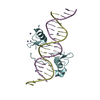
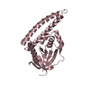
regulation of primitive erythrocyte differentiation / basophil differentiation / eosinophil fate commitment / regulation of definitive erythrocyte differentiation / regulation of glycoprotein biosynthetic process / megakaryocyte differentiation / RUNX1 regulates genes involved in megakaryocyte differentiation and platelet function / cell development / RUNX1 regulates transcription of genes involved in differentiation of HSCs / Sertoli cell development ...regulation of primitive erythrocyte differentiation / basophil differentiation / eosinophil fate commitment / regulation of definitive erythrocyte differentiation / regulation of glycoprotein biosynthetic process / megakaryocyte differentiation / RUNX1 regulates genes involved in megakaryocyte differentiation and platelet function / cell development / RUNX1 regulates transcription of genes involved in differentiation of HSCs / Sertoli cell development / dendritic cell differentiation / Factors involved in megakaryocyte development and platelet production / positive regulation of mast cell degranulation / cellular response to follicle-stimulating hormone stimulus / negative regulation of bone mineralization / myeloid cell differentiation / C2H2 zinc finger domain binding / embryonic hemopoiesis / platelet formation / DNA binding, bending / positive regulation of osteoblast proliferation / animal organ regeneration / negative regulation of extrinsic apoptotic signaling pathway in absence of ligand / cell fate commitment / cis-regulatory region sequence-specific DNA binding / erythrocyte development / transcription repressor complex / positive regulation of erythrocyte differentiation / homeostasis of number of cells within a tissue / cellular response to cAMP / transcription coregulator binding / erythrocyte differentiation / RNA polymerase II transcription regulatory region sequence-specific DNA binding / protein-DNA complex / chromatin DNA binding / platelet aggregation / male gonad development / transcription coactivator binding / sequence-specific double-stranded DNA binding / p53 binding / cell-cell signaling / cellular response to lipopolysaccharide / positive regulation of cytosolic calcium ion concentration / DNA-binding transcription activator activity, RNA polymerase II-specific / transcription regulator complex / sequence-specific DNA binding / in utero embryonic development / RNA polymerase II-specific DNA-binding transcription factor binding / DNA-binding transcription factor activity, RNA polymerase II-specific / transcription cis-regulatory region binding / RNA polymerase II cis-regulatory region sequence-specific DNA binding / DNA-binding transcription factor activity / negative regulation of cell population proliferation / chromatin binding / negative regulation of apoptotic process / positive regulation of DNA-templated transcription / negative regulation of transcription by RNA polymerase II / positive regulation of transcription by RNA polymerase II / DNA binding / zinc ion binding / nucleoplasm / nucleus Similarity search - Function
In the structure databanks used in Yorodumi, some data are registered as the other names, "COVID-19 virus" and "2019-nCoV". Here are the details of the virus and the list of structure data.
Jan 31, 2019. EMDB accession codes are about to change! (news from PDBe EMDB page)
EMDB accession codes are about to change! (news from PDBe EMDB page)
The allocation of 4 digits for EMDB accession codes will soon come to an end. Whilst these codes will remain in use, new EMDB accession codes will include an additional digit and will expand incrementally as the available range of codes is exhausted. The current 4-digit format prefixed with “EMD-” (i.e. EMD-XXXX) will advance to a 5-digit format (i.e. EMD-XXXXX), and so on. It is currently estimated that the 4-digit codes will be depleted around Spring 2019, at which point the 5-digit format will come into force.
The EM Navigator/Yorodumi systems omit the EMD- prefix.
Related info.:Q: What is EMD? / ID/Accession-code notation in Yorodumi/EM Navigator
Yorodumi is a browser for structure data from EMDB, PDB, SASBDB, etc.
This page is also the successor to EM Navigator detail page, and also detail information page/front-end page for Omokage search.
The word "yorodu" (or yorozu) is an old Japanese word meaning "ten thousand". "mi" (miru) is to see.
Related info.:EMDB / PDB / SASBDB / Comparison of 3 databanks / Yorodumi Search / Aug 31, 2016. New EM Navigator & Yorodumi / Yorodumi Papers / Jmol/JSmol / Function and homology information / Changes in new EM Navigator and Yorodumi
 Movie
Movie Controller

 Yorodumi
 Open data
 Basic information
 Components
 Keywords
 Function and homology information
 X-RAY DIFFRACTION /
  Authors
 Citation
 Structure visualization
 Downloads & links
 Other downloads


 PDBj
PDBj


 Assembly


 Mass: 59.044 Da / Num. of mol.: 1 / Source method: obtained synthetically / Formula: C2H3O2
 Mass: 65.409 Da / Num. of mol.: 3 / Source method: obtained synthetically / Formula: Zn
 Sample preparation
 Processing