 Movie
Movie Controller


+ Open data

- Basic information
| Entry | Database: PDB / ID: 3val | ||||||
|---|---|---|---|---|---|---|---|
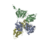
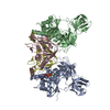
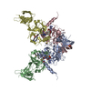
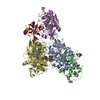
| Title | Structure of U2AF65 variant with BrU5C1 DNA | ||||||
 Components Components |
| ||||||
 Keywords Keywords | RNA BINDING PROTEIN/DNA / RNA SPLICING FACTOR / RNA RECOGNITION MOTIF / RNA BINDING PROTEIN / RNA BINDING PROTEIN-DNA complex | ||||||
| Function / homology |  Function and homology information Function and homology informationU2AF complex / poly-pyrimidine tract binding / pre-mRNA 3'-splice site binding / C2H2 zinc finger domain binding / mRNA 3'-end processing / commitment complex / Transport of Mature mRNA derived from an Intron-Containing Transcript / RNA Polymerase II Transcription Termination / U2-type prespliceosome / molecular function inhibitor activity ...U2AF complex / poly-pyrimidine tract binding / pre-mRNA 3'-splice site binding / C2H2 zinc finger domain binding / mRNA 3'-end processing / commitment complex / Transport of Mature mRNA derived from an Intron-Containing Transcript / RNA Polymerase II Transcription Termination / U2-type prespliceosome / molecular function inhibitor activity / spliceosomal complex assembly / Protein hydroxylation / negative regulation of mRNA splicing, via spliceosome / mRNA Polyadenylation / mRNA Splicing - Major Pathway / negative regulation of protein ubiquitination / positive regulation of RNA splicing / Dengue Virus-Host Interactions / spliceosomal complex / mRNA splicing, via spliceosome / mRNA processing / nuclear speck / enzyme binding / RNA binding / nucleoplasm / nucleus Similarity search - Function | ||||||
| Biological species |  Homo sapiens (human) Homo sapiens (human) | ||||||
| Method |  X-RAY DIFFRACTION / SYNCHROTRON / MOLECULAR REPLACEMENT / Resolution: 2.5 Å X-RAY DIFFRACTION / SYNCHROTRON / MOLECULAR REPLACEMENT / Resolution: 2.5 Å | ||||||
 Authors Authors | Jenkins, J.L. / Frato, K.H. / Kielkopf, C.L. | ||||||
 Citation Citation | Journal: Nucleic Acids Res. / Year: 2013 Title: U2AF65 adapts to diverse pre-mRNA splice sites through conformational selection of specific and promiscuous RNA recognition motifs. Authors: Jenkins, J.L. / Agrawal, A.A. / Gupta, A. / Green, M.R. / Kielkopf, C.L. | ||||||
| History |
|
- Structure visualization
| Structure viewer | Molecule: MolmilJmol/JSmol |
|---|
- Downloads & links
-Download
| PDBx/mmCIF format | 3val.cif.gz | 163.6 KB | Display | PDBx/mmCIF format |
|---|---|---|---|---|
| PDB format | pdb3val.ent.gz | 128.3 KB | Display | PDB format |
| PDBx/mmJSON format | 3val.json.gz | Tree view | PDBx/mmJSON format | |
| Others |  Other downloads Other downloads |
-Validation report
| Arichive directory | https://data.pdbj.org/pub/pdb/validation_reports/va/3valftp://data.pdbj.org/pub/pdb/validation_reports/va/3val | HTTPS FTP |
|---|
-Related structure data
| Related structure data |  3vafC  3vagC  3vahC  3vaiC  3vajC  3vakC  3vamC  2g4bS  3van 3vao S: Starting model for refinement C: citing same article ( |
|---|---|
| Similar structure data |


-Links
 PDBj
PDBj


- Assembly
| Deposited unit | 
| ||||||||
|---|---|---|---|---|---|---|---|---|---|
| 1 |
| ||||||||
| Unit cell |
|
-Components
| #1: Protein | Mass: 19075.754 Da / Num. of mol.: 4 / Fragment: RNA Binding Domains 1 and 2 Source method: isolated from a genetically manipulated source Source: (gene. exp.) Homo sapiens (human) / Gene: U2AF2, U2AF65 / Plasmid: PGEX-6P / Production host:  #2: DNA chain | Mass: 2064.121 Da / Num. of mol.: 4 / Source method: obtained synthetically / Details: DNA #3: Chemical | ChemComp-DIO /   Mass: 88.105 Da / Num. of mol.: 4 / Source method: obtained synthetically / Formula: C4H8O2 Mass: 88.105 Da / Num. of mol.: 4 / Source method: obtained synthetically / Formula: C4H8O2#4: Chemical | ChemComp-SO4 /   Mass: 96.063 Da / Num. of mol.: 11 / Source method: obtained synthetically / Formula: SO4 Mass: 96.063 Da / Num. of mol.: 11 / Source method: obtained synthetically / Formula: SO4#5: Water | ChemComp-HOH / |  Mass: 18.015 Da / Num. of mol.: 225 / Source method: isolated from a natural source / Formula: H2O Mass: 18.015 Da / Num. of mol.: 225 / Source method: isolated from a natural source / Formula: H2O |
|---|
-Experimental details
-Experiment
| Experiment | Method: X-RAY DIFFRACTION / Number of used crystals: 1 |
|---|
- Sample preparation
| Crystal | Density Matthews: 2.49 Å3/Da / Density % sol: 50.65 % |
|---|---|
| Crystal grow | Temperature: 277 K / Method: vapor diffusion, hanging drop / pH: 6.5 Details: 1.6M Ammonium sulfate, 10% Dioxane, 0.1M MES pH 6.5, VAPOR DIFFUSION, HANGING DROP, temperature 277K |
-Data collection
| Diffraction | Mean temperature: 100 K | |||||||||||||||||||||||||||||||||||||||||||||||||||||||||||||||||||||||||||||
|---|---|---|---|---|---|---|---|---|---|---|---|---|---|---|---|---|---|---|---|---|---|---|---|---|---|---|---|---|---|---|---|---|---|---|---|---|---|---|---|---|---|---|---|---|---|---|---|---|---|---|---|---|---|---|---|---|---|---|---|---|---|---|---|---|---|---|---|---|---|---|---|---|---|---|---|---|---|---|
| Diffraction source | Source: SYNCHROTRON / Site: NSLS  / Beamline: X29A / Wavelength: 0.912 Å / Beamline: X29A / Wavelength: 0.912 Å | |||||||||||||||||||||||||||||||||||||||||||||||||||||||||||||||||||||||||||||
| Detector | Type: ADSC QUANTUM 315 / Detector: CCD / Date: Jan 1, 2006 | |||||||||||||||||||||||||||||||||||||||||||||||||||||||||||||||||||||||||||||
| Radiation | Protocol: SINGLE WAVELENGTH / Monochromatic (M) / Laue (L): M / Scattering type: x-ray | |||||||||||||||||||||||||||||||||||||||||||||||||||||||||||||||||||||||||||||
| Radiation wavelength | Wavelength: 0.912 Å / Relative weight: 1 | |||||||||||||||||||||||||||||||||||||||||||||||||||||||||||||||||||||||||||||
| Reflection | Resolution: 2.5→50 Å / % possible obs: 95.1 % / Observed criterion σ(I): -1 / Redundancy: 6.7 % / Rmerge(I) obs: 0.147 / Χ2: 1.635 / Net I/σ(I): 17.7 | |||||||||||||||||||||||||||||||||||||||||||||||||||||||||||||||||||||||||||||
| Reflection shell |
|
- Processing
| Software |
| ||||||||||||||||||||||||||||
|---|---|---|---|---|---|---|---|---|---|---|---|---|---|---|---|---|---|---|---|---|---|---|---|---|---|---|---|---|---|
| Refinement | Method to determine structure: MOLECULAR REPLACEMENT Starting model: 2G4B Resolution: 2.5→50 Å / Occupancy max: 1 / Occupancy min: 1 / Cross valid method: THROUGHOUT / σ(F): 0 / Stereochemistry target values: Engh & Huber
| ||||||||||||||||||||||||||||
| Solvent computation | Bsol: 38.3339 Å2 | ||||||||||||||||||||||||||||
| Displacement parameters | Biso max: 123.62 Å2 / Biso mean: 39.7133 Å2 / Biso min: 10.82 Å2
| ||||||||||||||||||||||||||||
| Refinement step | Cycle: LAST / Resolution: 2.5→50 Å
| ||||||||||||||||||||||||||||
| Refine LS restraints |
| ||||||||||||||||||||||||||||
| Xplor file |
|
