 Movie
Movie Controller


+ Open data

- Basic information
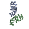
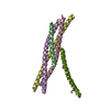
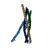
| Entry | Database: PDB / ID: 2g4b | ||||||
|---|---|---|---|---|---|---|---|
| Title | Structure of U2AF65 variant with polyuridine tract | ||||||
 Components Components |
| ||||||
 Keywords Keywords | RNA Binding Protein/RNA / protein-RNA complex / RNA splicing factor / RNA recognition motif / RNA Binding Protein-RNA COMPLEX | ||||||
| Function / homology |  Function and homology information Function and homology informationU2AF complex / poly-pyrimidine tract binding / pre-mRNA 3'-splice site binding / C2H2 zinc finger domain binding / mRNA 3'-end processing / commitment complex / Transport of Mature mRNA derived from an Intron-Containing Transcript / RNA Polymerase II Transcription Termination / U2-type prespliceosome / molecular function inhibitor activity ...U2AF complex / poly-pyrimidine tract binding / pre-mRNA 3'-splice site binding / C2H2 zinc finger domain binding / mRNA 3'-end processing / commitment complex / Transport of Mature mRNA derived from an Intron-Containing Transcript / RNA Polymerase II Transcription Termination / U2-type prespliceosome / molecular function inhibitor activity / negative regulation of mRNA splicing, via spliceosome / spliceosomal complex assembly / Protein hydroxylation / mRNA Splicing - Major Pathway / negative regulation of protein ubiquitination / positive regulation of RNA splicing / spliceosomal complex / mRNA splicing, via spliceosome / mRNA processing / nuclear speck / enzyme binding / RNA binding / nucleoplasm / nucleus Similarity search - Function | ||||||
| Biological species |  Homo sapiens (human) Homo sapiens (human) | ||||||
| Method |  X-RAY DIFFRACTION / SYNCHROTRON / MOLECULAR REPLACEMENT / Resolution: 2.5 Å X-RAY DIFFRACTION / SYNCHROTRON / MOLECULAR REPLACEMENT / Resolution: 2.5 Å | ||||||
 Authors Authors | Sickmier, E.A. / Kielkopf, C.L. | ||||||
 Citation Citation | Journal: Mol.Cell / Year: 2006 Title: Structural basis of polypyrimidine tract recognition by the essential splicing factor U2AF65. Authors: Sickmier, E.A. / Frato, K.E. / Paranawithana, S. / Shen, H. / Green, M.R. / Kielkopf, C.L. #1: Journal: Acta Crystallogr.,Sect.F / Year: 2006 Title: Crystallization and preliminary X-ray analysis of a U2AF65 variant in complex with a polypyrimidine-tract analogue by use of protein engineering. Authors: Sickmier, E.A. / Frato, K.E. / Kielkopf, C.L. | ||||||
| History |
|
- Structure visualization
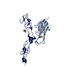
| Structure viewer | Molecule: MolmilJmol/JSmol |
|---|
- Downloads & links
-Download
| PDBx/mmCIF format | 2g4b.cif.gz | 51.4 KB | Display | PDBx/mmCIF format |
|---|---|---|---|---|
| PDB format | pdb2g4b.ent.gz | 36.1 KB | Display | PDB format |
| PDBx/mmJSON format | 2g4b.json.gz | Tree view | PDBx/mmJSON format | |
| Others |  Other downloads Other downloads |
-Validation report
| Arichive directory | https://data.pdbj.org/pub/pdb/validation_reports/g4/2g4bftp://data.pdbj.org/pub/pdb/validation_reports/g4/2g4b | HTTPS FTP |
|---|
-Related structure data
| Related structure data |  2fzr S: Starting model for refinement |
|---|---|
| Similar structure data |


-Links
 PDBj
PDBj


- Assembly
| Deposited unit | 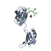
| ||||||||
|---|---|---|---|---|---|---|---|---|---|
| 1 | 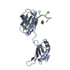
| ||||||||
| Unit cell |
| ||||||||
| Details | The second part of the biological assembly is generated by the following six-fold axis: MATRIX 0.5001393 0.8659449 0.0003453 -0.8659446 0.5001394 -0.0007568 -0.0008280 0.0000795 0.9999996 TRANSLATION 129.3170471 -0.0240173 -10.3325310 |
-Components
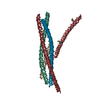
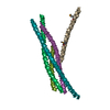
| #1: RNA chain | Mass: 2098.203 Da / Num. of mol.: 1 / Source method: obtained synthetically / Details: polypyrimidine tract |
|---|---|
| #2: Protein | Mass: 18921.586 Da / Num. of mol.: 1 / Fragment: RNA binding domain / Mutation: deletion of residues 238-258 Source method: isolated from a genetically manipulated source Source: (gene. exp.) Homo sapiens (human) / Gene: U2AF65 / Plasmid: pGEX-6p / Species (production host): Escherichia coli / Production host:  |
| #3: Chemical | ChemComp-DIO /   Mass: 88.105 Da / Num. of mol.: 1 / Source method: obtained synthetically / Formula: C4H8O2 Mass: 88.105 Da / Num. of mol.: 1 / Source method: obtained synthetically / Formula: C4H8O2 |
| #4: Water | ChemComp-HOH /  Mass: 18.015 Da / Num. of mol.: 30 / Source method: isolated from a natural source / Formula: H2O Mass: 18.015 Da / Num. of mol.: 30 / Source method: isolated from a natural source / Formula: H2O |
-Experimental details
-Experiment
| Experiment | Method: X-RAY DIFFRACTION / Number of used crystals: 1 |
|---|
- Sample preparation
| Crystal | Density Matthews: 3.6 Å3/Da / Density % sol: 65.83 % | ||||||||||||||||||||||||||||||||||||||||||||||||
|---|---|---|---|---|---|---|---|---|---|---|---|---|---|---|---|---|---|---|---|---|---|---|---|---|---|---|---|---|---|---|---|---|---|---|---|---|---|---|---|---|---|---|---|---|---|---|---|---|---|
| Crystal grow | Temperature: 277 K / Method: vapor diffusion, hanging drop / pH: 6.5 Details: .6 M ammonium sulfate, 100 mM 2-(N-morpholino) ethanesulfonic acid, sodium salt (MES) pH 6.5, 10% dioxane, 200 mM non-detergent sulfobetaine (NDSB) 195 , VAPOR DIFFUSION, HANGING DROP, temperature 277K | ||||||||||||||||||||||||||||||||||||||||||||||||
| Components of the solutions |
|
-Data collection
| Diffraction | Mean temperature: 100 K |
|---|---|
| Diffraction source | Source: SYNCHROTRON / Site: NSLS  / Beamline: X8C / Wavelength: 1 Å / Beamline: X8C / Wavelength: 1 Å |
| Detector | Type: ADSC QUANTUM 4 / Detector: CCD / Date: Apr 6, 2005 |
| Radiation | Monochromator: parabolic collimating mirror placed upstream of the monochromator Protocol: SINGLE WAVELENGTH / Monochromatic (M) / Laue (L): M / Scattering type: x-ray |
| Radiation wavelength | Wavelength: 1 Å / Relative weight: 1 |
| Reflection | Resolution: 2.5→20 Å / Num. all: 11182 / Num. obs: 11182 / % possible obs: 99.7 % / Observed criterion σ(F): -1 / Observed criterion σ(I): -1 |
| Reflection shell | Resolution: 2.5→2.59 Å / % possible all: 99.7 |
- Processing
| Software |
| ||||||||||||||||||||
|---|---|---|---|---|---|---|---|---|---|---|---|---|---|---|---|---|---|---|---|---|---|
| Refinement | Method to determine structure: MOLECULAR REPLACEMENT Starting model: PDB Entry: 2FZR 2fzr Resolution: 2.5→20 Å / σ(F): 0 / Stereochemistry target values: Engh & Huber
| ||||||||||||||||||||
| Refinement step | Cycle: LAST / Resolution: 2.5→20 Å
| ||||||||||||||||||||
| Refine LS restraints |
|
