 Movie
Movie Controller


+ Open data

- Basic information
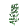
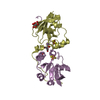
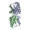
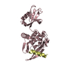
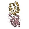
| Entry | Database: PDB / ID: 2db0 | ||||||
|---|---|---|---|---|---|---|---|
| Title | Crystal structure of PH0542 | ||||||
 Components Components | 253aa long hypothetical protein | ||||||
 Keywords Keywords | PROTEIN BINDING / HEAT repeats / helical structure / Structural Genomics / NPPSFA / National Project on Protein Structural and Functional Analyses / RIKEN Structural Genomics/Proteomics Initiative / RSGI | ||||||
| Function / homology |  Function and homology information Function and homology informationRepeat of uncharacterized protein PH0542 / Repeat of uncharacterized protein PH0542 / HEAT repeat / HEAT repeat / HEAT, type 2 / HEAT repeat profile. / Leucine-rich Repeat Variant / Leucine-rich Repeat Variant / Alpha Horseshoe / Armadillo-like helical ...Repeat of uncharacterized protein PH0542 / Repeat of uncharacterized protein PH0542 / HEAT repeat / HEAT repeat / HEAT, type 2 / HEAT repeat profile. / Leucine-rich Repeat Variant / Leucine-rich Repeat Variant / Alpha Horseshoe / Armadillo-like helical / Armadillo-type fold / Mainly Alpha Similarity search - Domain/homology | ||||||
| Biological species |   Pyrococcus horikoshii (archaea) Pyrococcus horikoshii (archaea) | ||||||
| Method |  X-RAY DIFFRACTION / SYNCHROTRON / MAD / Resolution: 2.2 Å X-RAY DIFFRACTION / SYNCHROTRON / MAD / Resolution: 2.2 Å | ||||||
 Authors Authors | Nishino, A. / Handa, N. / Kishishita, S. / Murayama, K. / Shirouzu, M. / RIKEN Structural Genomics/Proteomics Initiative (RSGI) | ||||||
 Citation Citation | Journal: To be Published Title: Crystal structure of PH0542 Authors: Handa, N. / Nishino, A. / Kishishita, S. / Murayama, K. / Shirouzu, M. / Yokoyama, S. | ||||||
| History |
|
- Structure visualization
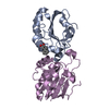
| Structure viewer | Molecule: MolmilJmol/JSmol |
|---|
- Downloads & links
-Download
| PDBx/mmCIF format | 2db0.cif.gz | 105.4 KB | Display | PDBx/mmCIF format |
|---|---|---|---|---|
| PDB format | pdb2db0.ent.gz | 83 KB | Display | PDB format |
| PDBx/mmJSON format | 2db0.json.gz | Tree view | PDBx/mmJSON format | |
| Others |  Other downloads Other downloads |
-Validation report
| Arichive directory | https://data.pdbj.org/pub/pdb/validation_reports/db/2db0ftp://data.pdbj.org/pub/pdb/validation_reports/db/2db0 | HTTPS FTP |
|---|
-Related structure data
| Similar structure data | |
|---|---|
| Other databases |


-Links
 PDBj
PDBj
- Assembly
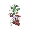
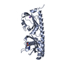
| Deposited unit | 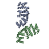
| ||||||||
|---|---|---|---|---|---|---|---|---|---|
| 1 | 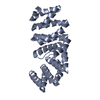
| ||||||||
| 2 | 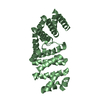
| ||||||||
| Unit cell |
|
-Components
| #1: Protein | Mass: 28734.666 Da / Num. of mol.: 2 Source method: isolated from a genetically manipulated source Source: (gene. exp.) Pyrococcus horikoshii (archaea) / Strain: OT3 / Plasmid: pET11a / Production host:  #2: Water | ChemComp-HOH / |  Mass: 18.015 Da / Num. of mol.: 101 / Source method: isolated from a natural source / Formula: H2O Mass: 18.015 Da / Num. of mol.: 101 / Source method: isolated from a natural source / Formula: H2O |
|---|
-Experimental details
-Experiment
| Experiment | Method: X-RAY DIFFRACTION / Number of used crystals: 1 |
|---|
- Sample preparation
| Crystal | Density Matthews: 2.07 Å3/Da / Density % sol: 40.5 % |
|---|---|
| Crystal grow | Temperature: 293 K / Method: vapor diffusion, hanging drop / pH: 8.5 Details: 20% PEG3350, 0.2M ammonium acetate, 0.1M Tris-HCl, pH 8.5, VAPOR DIFFUSION, HANGING DROP, temperature 293K |
-Data collection
| Diffraction | Mean temperature: 100 K | ||||||||||||
|---|---|---|---|---|---|---|---|---|---|---|---|---|---|
| Diffraction source | Source: SYNCHROTRON / Site: SPring-8  / Beamline: BL44B2 / Wavelength: 0.9790, 0.9797, 0.9670 / Beamline: BL44B2 / Wavelength: 0.9790, 0.9797, 0.9670 | ||||||||||||
| Detector | Type: MARRESEARCH / Detector: CCD / Date: Jun 20, 2005 / Details: mirrors | ||||||||||||
| Radiation | Monochromator: Si / Protocol: MAD / Monochromatic (M) / Laue (L): M / Scattering type: x-ray | ||||||||||||
| Radiation wavelength |
| ||||||||||||
| Reflection | Resolution: 2.2→50 Å / Num. obs: 24884 / % possible obs: 99.3 % / Observed criterion σ(I): -3 / Redundancy: 6 % / Rsym value: 0.075 / Net I/σ(I): 17 | ||||||||||||
| Reflection shell | Resolution: 2.2→2.28 Å / Mean I/σ(I) obs: 4.6 / Rsym value: 0.369 / % possible all: 98.8 |
- Processing
| Software |
| ||||||||||||||||||||
|---|---|---|---|---|---|---|---|---|---|---|---|---|---|---|---|---|---|---|---|---|---|
| Refinement | Method to determine structure: MAD / Resolution: 2.2→50 Å / Cross valid method: THROUGHOUT / Stereochemistry target values: MAXIMUM LIKELIHOOD / Details: This structure was refined also by CNS 1.1.
| ||||||||||||||||||||
| Displacement parameters | Biso mean: 36.6 Å2
| ||||||||||||||||||||
| Refinement step | Cycle: LAST / Resolution: 2.2→50 Å
| ||||||||||||||||||||
| Refine LS restraints |
| ||||||||||||||||||||
| LS refinement shell | Resolution: 2.2→2.257 Å
|
