Mass: 18.015 Da / Num. of mol.: 11 / Source method: isolated from a natural source / Formula: H2O
Has protein modification
Y
Sequence details
THE AUTHORS STATE THAT THE SEQUENCE "NV" IS THE ORIGINAL SEQUENCE FROM ENDOTHELIUM, AS OPPOSED TO ...THE AUTHORS STATE THAT THE SEQUENCE "NV" IS THE ORIGINAL SEQUENCE FROM ENDOTHELIUM, AS OPPOSED TO KSL WHICH IS THE GENOMIC SEQUENCE. THE UNIPROT RECORD WAS CHANGED TO THE GENOMIC SEQUENCE "KSL" AFTER THE CLONE WAS CREATED AND THE CONSTRUCT WAS NOT CHANGED BECAUSE IT WAS PERFORMING WELL.
-
Experimental details
-
Experiment
Experiment
Method: X-RAY DIFFRACTION / Number of used crystals: 77
-
Sample preparation
Crystal
Density Matthews: 2.61 Å3/Da / Density % sol: 52.93 %
Crystal grow
Temperature: 287 K / Method: lipidic cubic phase Details: 0.1M Tricine, 34-36% PEG400, 80mM sodium citrate and 4% glycerol, Lipid cubic phase, temperature 287K
-
Data collection
Diffraction
ID
Mean temperature (K)
Crystal-ID
1
100
1
2
1
Diffraction source
Source
Site
Beamline
ID
SYNCHROTRON
APS
23-ID-B
1
SYNCHROTRON
APS
23-ID-D
2
Detector
Type: MARMOSAIC 300 mm CCD / Detector: CCD / Date: Jan 1, 2009
Radiation
Protocol: SINGLE WAVELENGTH / Monochromatic (M) / Laue (L): M / Scattering type: x-ray
Radiation wavelength
Relative weight: 1
Reflection
Resolution: 2.8→20 Å / Num. obs: 15297 / % possible obs: 97.2 % / Redundancy: 6 % / Biso Wilson estimate: 34.3 Å2 / Rmerge(I) obs: 0.16 / Net I/σ(I): 6.1
Reflection shell
Resolution: 2.8→2.95 Å / Redundancy: 3.1 % / Rmerge(I) obs: 0.42 / Mean I/σ(I) obs: 1.9 / % possible all: 94.4
-
Processing
Software
Name
Version
Classification
PHASER
phasing
BUSTER
2.8.0
refinement
XDS
datareduction
Microdiffaction
dataassemblymethods
datascaling
SCALA
datascaling
Refinement
Method to determine structure: MOLECULAR REPLACEMENT Starting model: 7TM of b2AR and T4L Resolution: 2.8→19.52 Å / Cor.coef. Fo:Fc: 0.9055 / Cor.coef. Fo:Fc free: 0.8586 / Cross valid method: THROUGHOUT / σ(F): 0
Rfactor
Num. reflection
% reflection
Selection details
Rfree
0.2717
742
4.86 %
RANDOM
Rwork
0.2287
-
-
-
obs
0.2307
15275
-
-
Displacement parameters
Biso mean: 76.61 Å2
Baniso -1
Baniso -2
Baniso -3
1-
7.5487 Å2
0 Å2
0 Å2
2-
-
1.2749 Å2
0 Å2
3-
-
-
-8.8236 Å2
Refine analyze
Luzzati coordinate error obs: 0.44 Å
Refinement step
Cycle: LAST / Resolution: 2.8→19.52 Å
Protein
Nucleic acid
Ligand
Solvent
Total
Num. atoms
3475
0
37
11
3523
Refine LS restraints
Refine-ID
Type
Dev ideal
Number
Restraint function
Weight
X-RAY DIFFRACTION
t_bond_d
0.01
3585
HARMONIC
2
X-RAY DIFFRACTION
t_angle_deg
0.65
4893
HARMONIC
3
X-RAY DIFFRACTION
t_dihedral_angle_d
1597
SINUSOIDAL
20
X-RAY DIFFRACTION
t_incorr_chiral_ct
X-RAY DIFFRACTION
t_pseud_angle
X-RAY DIFFRACTION
t_trig_c_planes
56
HARMONIC
2
X-RAY DIFFRACTION
t_gen_planes
529
HARMONIC
5
X-RAY DIFFRACTION
t_it
3585
HARMONIC
20
X-RAY DIFFRACTION
t_nbd
0
SEMIHARMONIC
5
X-RAY DIFFRACTION
t_omega_torsion
2.24
X-RAY DIFFRACTION
t_other_torsion
2.2
X-RAY DIFFRACTION
t_improper_torsion
X-RAY DIFFRACTION
t_chiral_improper_torsion
511
SEMIHARMONIC
5
X-RAY DIFFRACTION
t_sum_occupancies
X-RAY DIFFRACTION
t_utility_distance
X-RAY DIFFRACTION
t_utility_angle
X-RAY DIFFRACTION
t_utility_torsion
X-RAY DIFFRACTION
t_ideal_dist_contact
4387
SEMIHARMONIC
4
LS refinement shell
Resolution: 2.8→2.99 Å / Total num. of bins used: 8
Rfactor
Num. reflection
% reflection
Rfree
0.2762
125
4.72 %
Rwork
0.2596
2522
-
all
0.2604
2647
-
Refinement TLS params.
Method: refined / Origin x: 17.1445 Å / Origin y: 18.5316 Å / Origin z: 13.5414 Å
11
12
13
21
22
23
31
32
33
T
0.2273 Å2
0.0573 Å2
-0.0048 Å2
-
-0.0572 Å2
0.0225 Å2
-
-
-0.1172 Å2
L
1.7528 °2
-0.2526 °2
0.7309 °2
-
0 °2
-0.5602 °2
-
-
0.4655 °2
S
0.0421 Å °
-0.2737 Å °
-0.1428 Å °
-0.1301 Å °
-0.0121 Å °
-0.0197 Å °
0.0116 Å °
-0.0999 Å °
-0.03 Å °
Refinement TLS group
Selection details: chain A
+
About Yorodumi
-
News
-
Feb 9, 2022. New format data for meta-information of EMDB entries
New format data for meta-information of EMDB entries
Version 3 of the EMDB header file is now the official format.
The previous official version 1.9 will be removed from the archive.
In the structure databanks used in Yorodumi, some data are registered as the other names, "COVID-19 virus" and "2019-nCoV". Here are the details of the virus and the list of structure data.
Jan 31, 2019. EMDB accession codes are about to change! (news from PDBe EMDB page)
EMDB accession codes are about to change! (news from PDBe EMDB page)
The allocation of 4 digits for EMDB accession codes will soon come to an end. Whilst these codes will remain in use, new EMDB accession codes will include an additional digit and will expand incrementally as the available range of codes is exhausted. The current 4-digit format prefixed with “EMD-” (i.e. EMD-XXXX) will advance to a 5-digit format (i.e. EMD-XXXXX), and so on. It is currently estimated that the 4-digit codes will be depleted around Spring 2019, at which point the 5-digit format will come into force.
The EM Navigator/Yorodumi systems omit the EMD- prefix.
Related info.:Q: What is EMD? / ID/Accession-code notation in Yorodumi/EM Navigator
Yorodumi is a browser for structure data from EMDB, PDB, SASBDB, etc.
This page is also the successor to EM Navigator detail page, and also detail information page/front-end page for Omokage search.
The word "yorodu" (or yorozu) is an old Japanese word meaning "ten thousand". "mi" (miru) is to see.
Related info.:EMDB / PDB / SASBDB / Comparison of 3 databanks / Yorodumi Search / Aug 31, 2016. New EM Navigator & Yorodumi / Yorodumi Papers / Jmol/JSmol / Function and homology information / Changes in new EM Navigator and Yorodumi
 Movie
Movie Controller


 Open data
 Basic information
 Components
 Keywords
 Function and homology information
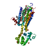
 Homo sapiens (human)
 Enterobacteria phage T4 (virus)
 X-RAY DIFFRACTION /
  Authors
 Citation
 Structure visualization
 Downloads & links
 Other downloads
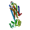


 PDBj
PDBj


 Assembly

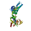
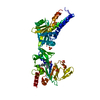
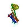
 Spodoptera frugiperda (fall armyworm) / References: UniProt: P21453, UniProt: P00720, lysozyme
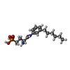
 Mass: 342.370 Da / Num. of mol.: 1 / Source method: obtained synthetically / Formula: C16H27N2O4P
 Type: D-saccharide, beta linking / Mass: 221.208 Da / Num. of mol.: 1
 Mass: 18.015 Da / Num. of mol.: 11 / Source method: isolated from a natural source / Formula: H2O
 Sample preparation
 Processing