 Movie
Movie Controller

[English] 日本語
 Yorodumi
- PDB-3uzb: Crystal Structures of Branched-Chain Aminotransferase from Deinoc... -
+ Open data

- Basic information
| Entry | Database: PDB / ID: 3uzb | ||||||
|---|---|---|---|---|---|---|---|
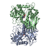
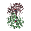
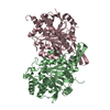
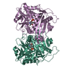
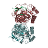
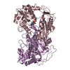
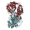
| Title | Crystal Structures of Branched-Chain Aminotransferase from Deinococcus radiodurans Complexes with alpha-Ketoisocaproate and L-Glutamate Suggest Its Radio-Resistance for Catalysis | ||||||
 Components Components | Branched-chain-amino-acid aminotransferase | ||||||
 Keywords Keywords | TRANSFERASE / BCAT / Amino-acid biosynthesis / Aminotransferase / Branched-chain amino acid biosynthesis / Pyridoxal phosphate / alpha-Ketoisocaproate | ||||||
| Function / homology |  Function and homology information Function and homology informationbranched-chain-amino-acid:2-oxoglutarate transaminase activity / L-valine:2-oxoglutarate transaminase activity / L-isoleucine:2-oxoglutarate transaminase activity / L-leucine:2-oxoglutarate transaminase activity / branched-chain-amino-acid transaminase / L-leucine biosynthetic process / L-valine biosynthetic process / : Similarity search - Function | ||||||
| Biological species |  Deinococcus radiodurans (radioresistant) Deinococcus radiodurans (radioresistant) | ||||||
| Method |  X-RAY DIFFRACTION / SYNCHROTRON / MOLECULAR REPLACEMENT / Resolution: 3 Å X-RAY DIFFRACTION / SYNCHROTRON / MOLECULAR REPLACEMENT / Resolution: 3 Å | ||||||
 Authors Authors | Chen, C.D. / Huang, Y.C. / Chuankhayan, P. / Hsieh, Y.C. / Huang, T.F. / Lin, C.H. / Guan, H.H. / Liu, M.Y. / Chang, W.C. / Chen, C.J. | ||||||
 Citation Citation | Journal: J.Bacteriol. / Year: 2012 Title: Crystal Structures of Complexes of the Branched-Chain Aminotransferase from Deinococcus radiodurans with alpha-Ketoisocaproate and L-Glutamate Suggest the Radiation Resistance of This Enzyme for Catalysis Authors: Chen, C.D. / Lin, C.H. / Chuankhayan, P. / Huang, Y.C. / Hsieh, Y.C. / Huang, T.F. / Guan, H.H. / Liu, M.Y. / Chang, W.C. / Chen, C.J. | ||||||
| History |
|
- Structure visualization
| Structure viewer | Molecule: MolmilJmol/JSmol |
|---|
- Downloads & links
-Download
| PDBx/mmCIF format | 3uzb.cif.gz | 266.3 KB | Display | PDBx/mmCIF format |
|---|---|---|---|---|
| PDB format | pdb3uzb.ent.gz | 215.2 KB | Display | PDB format |
| PDBx/mmJSON format | 3uzb.json.gz | Tree view | PDBx/mmJSON format | |
| Others |  Other downloads Other downloads |
-Validation report
| Arichive directory | https://data.pdbj.org/pub/pdb/validation_reports/uz/3uzbftp://data.pdbj.org/pub/pdb/validation_reports/uz/3uzb | HTTPS FTP |
|---|
-Related structure data
| Related structure data |  3uyyC  3uzoC  2cojS C: citing same article ( S: Starting model for refinement |
|---|---|
| Similar structure data |


-Links
 PDBj
PDBj
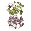
- Assembly
| Deposited unit | 
| ||||||||
|---|---|---|---|---|---|---|---|---|---|
| 1 | 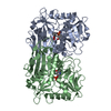
| ||||||||
| 2 | 
| ||||||||
| Unit cell |
|
-Components
| #1: Protein | Mass: 39465.324 Da / Num. of mol.: 4 Source method: isolated from a genetically manipulated source Source: (gene. exp.) Deinococcus radiodurans (radioresistant)Strain: R1 / Gene: DR_1626 / Plasmid: pQE30 / Production host: References: UniProt: Q9RTX5, branched-chain-amino-acid transaminase #2: Chemical | ChemComp-PLP /   Mass: 247.142 Da / Num. of mol.: 4 / Source method: obtained synthetically / Formula: C8H10NO6P Mass: 247.142 Da / Num. of mol.: 4 / Source method: obtained synthetically / Formula: C8H10NO6P#3: Chemical | ChemComp-COI / 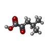  Mass: 130.142 Da / Num. of mol.: 4 / Source method: obtained synthetically / Formula: C6H10O3 Mass: 130.142 Da / Num. of mol.: 4 / Source method: obtained synthetically / Formula: C6H10O3#4: Water | ChemComp-HOH / |  Mass: 18.015 Da / Num. of mol.: 173 / Source method: isolated from a natural source / Formula: H2O Mass: 18.015 Da / Num. of mol.: 173 / Source method: isolated from a natural source / Formula: H2O |
|---|
-Experimental details
-Experiment
| Experiment | Method: X-RAY DIFFRACTION / Number of used crystals: 1 |
|---|
- Sample preparation
| Crystal | Density Matthews: 2.33 Å3/Da / Density % sol: 47.29 % |
|---|---|
| Crystal grow | Temperature: 293 K / Method: vapor diffusion, hanging drop / pH: 7.4 Details: 0.15M magnesium chloride, 15%(w/v) polyethylene glycol 8000, 0.1M Tris(pH 7.3), VAPOR DIFFUSION, HANGING DROP, temperature 293K |
-Data collection
| Diffraction | Mean temperature: 100 K |
|---|---|
| Diffraction source | Source: SYNCHROTRON / Site: NSRRC  / Beamline: BL13B1 / Wavelength: 1 Å / Beamline: BL13B1 / Wavelength: 1 Å |
| Detector | Type: ADSC QUANTUM 315r / Detector: CCD / Date: Nov 3, 2006 |
| Radiation | Monochromator: GRAPHITE / Protocol: SINGLE WAVELENGTH / Monochromatic (M) / Laue (L): M / Scattering type: x-ray |
| Radiation wavelength | Wavelength: 1 Å / Relative weight: 1 |
| Reflection | Resolution: 3→30 Å / Num. all: 34610 / Num. obs: 29754 / % possible obs: 87.7 % / Observed criterion σ(F): 2 / Observed criterion σ(I): 2 / Redundancy: 4.4 % / Biso Wilson estimate: 26.2 Å2 / Rmerge(I) obs: 0.12 / Rsym value: 0.14 / Net I/σ(I): 8.85 |
| Reflection shell | Resolution: 3→3.06 Å / Redundancy: 3.1 % / Rmerge(I) obs: 0.39 / Mean I/σ(I) obs: 2.46 / Num. unique all: 3370 / Rsym value: 0.417 / % possible all: 87.7 |
- Processing
| Software |
| |||||||||||||||||||||||||
|---|---|---|---|---|---|---|---|---|---|---|---|---|---|---|---|---|---|---|---|---|---|---|---|---|---|---|
| Refinement | Method to determine structure: MOLECULAR REPLACEMENT Starting model: PDB 2COJ Resolution: 3→30 Å / Cross valid method: THROUGHOUT / σ(F): 2 / Stereochemistry target values: Engh & Huber
| |||||||||||||||||||||||||
| Displacement parameters | Biso mean: 24.6723 Å2
| |||||||||||||||||||||||||
| Refinement step | Cycle: LAST / Resolution: 3→30 Å
| |||||||||||||||||||||||||
| Refine LS restraints |
| |||||||||||||||||||||||||
| LS refinement shell | Resolution: 3→3.06 Å / Rfactor Rfree error: 0.39
|
