 Movie
Movie Controller

[English] 日本語
 Yorodumi
- PDB-6l07: Crystal structure of Escherichia coli phosphatidylserine decarbox... -
+ Open data

- Basic information
| Entry | Database: PDB / ID: 6l07 | ||||||||||||
|---|---|---|---|---|---|---|---|---|---|---|---|---|---|
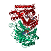
| Title | Crystal structure of Escherichia coli phosphatidylserine decarboxylase (PE-bound form) | ||||||||||||
 Components Components |
| ||||||||||||
 Keywords Keywords | LYASE / Phosphatidylserine / Phosphatidylethanolamine / Membrane | ||||||||||||
| Function / homology |  Function and homology information Function and homology informationphosphatidylserine decarboxylase / phosphatidylserine decarboxylase activity / phosphatidylethanolamine biosynthetic process / zymogen activation / protein autoprocessing / protein homodimerization activity / plasma membrane Similarity search - Function | ||||||||||||
| Biological species |  | ||||||||||||
| Method |  X-RAY DIFFRACTION / SYNCHROTRON / MOLECULAR REPLACEMENT / Resolution: 3.6 Å X-RAY DIFFRACTION / SYNCHROTRON / MOLECULAR REPLACEMENT / Resolution: 3.6 Å | ||||||||||||
 Authors Authors | Watanabe, Y. / Watanabe, S. | ||||||||||||
 Citation Citation | Journal: Structure / Year: 2020 Title: Structural Basis for Phosphatidylethanolamine Biosynthesis by Bacterial Phosphatidylserine Decarboxylase. Authors: Watanabe, Y. / Watanabe, Y. / Watanabe, S. | ||||||||||||
| History |
|
- Structure visualization
| Structure viewer | Molecule: MolmilJmol/JSmol |
|---|
- Downloads & links
-Download
| PDBx/mmCIF format | 6l07.cif.gz | 455.7 KB | Display | PDBx/mmCIF format |
|---|---|---|---|---|
| PDB format | pdb6l07.ent.gz | 371.7 KB | Display | PDB format |
| PDBx/mmJSON format | 6l07.json.gz | Tree view | PDBx/mmJSON format | |
| Others |  Other downloads Other downloads |
-Validation report
| Arichive directory | https://data.pdbj.org/pub/pdb/validation_reports/l0/6l07ftp://data.pdbj.org/pub/pdb/validation_reports/l0/6l07 | HTTPS FTP |
|---|
-Related structure data


-Links
 PDBj
PDBj
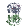
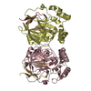
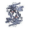
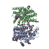
- Assembly
| Deposited unit | 
| ||||||||
|---|---|---|---|---|---|---|---|---|---|
| 1 | 
| ||||||||
| 2 | 
| ||||||||
| 3 | 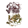
| ||||||||
| 4 | 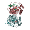
| ||||||||
| Unit cell |
|
-Components
| #1: Protein | Mass: 30263.859 Da / Num. of mol.: 8 Source method: isolated from a genetically manipulated source Source: (gene. exp.) References: UniProt: A0A446DLT6, UniProt: P0A8K1*PLUS, phosphatidylserine decarboxylase #2: Protein/peptide | Mass: 3755.313 Da / Num. of mol.: 5 Source method: isolated from a genetically manipulated source Source: (gene. exp.) References: UniProt: A0A446DLT6, UniProt: P0A8K1*PLUS, phosphatidylserine decarboxylase #3: Protein/peptide | Mass: 3769.296 Da / Num. of mol.: 3 Source method: isolated from a genetically manipulated source Source: (gene. exp.) References: UniProt: A0A446DLT6, UniProt: P0A8K1*PLUS, phosphatidylserine decarboxylase #4: Chemical | ChemComp-PEE /   Mass: 744.034 Da / Num. of mol.: 5 / Source method: obtained synthetically / Formula: C41H78NO8P / Comment: DOPE, phospholipid*YM Mass: 744.034 Da / Num. of mol.: 5 / Source method: obtained synthetically / Formula: C41H78NO8P / Comment: DOPE, phospholipid*YMHas ligand of interest | Y | |
|---|
-Experimental details
-Experiment
| Experiment | Method: X-RAY DIFFRACTION / Number of used crystals: 1 |
|---|
- Sample preparation
| Crystal | Density Matthews: 3.63 Å3/Da / Density % sol: 66.11 % |
|---|---|
| Crystal grow | Temperature: 293 K / Method: vapor diffusion, sitting drop / pH: 6.5 / Details: pentaerythritol ethoxylate, ammonium sulfate |
-Data collection
| Diffraction | Mean temperature: 100 K / Serial crystal experiment: N |
|---|---|
| Diffraction source | Source: SYNCHROTRON / Site: SPring-8  / Beamline: BL41XU / Wavelength: 1 Å / Beamline: BL41XU / Wavelength: 1 Å |
| Detector | Type: DECTRIS EIGER X 16M / Detector: PIXEL / Date: Apr 21, 2019 |
| Radiation | Protocol: SINGLE WAVELENGTH / Monochromatic (M) / Laue (L): M / Scattering type: x-ray |
| Radiation wavelength | Wavelength: 1 Å / Relative weight: 1 |
| Reflection | Resolution: 3.6→50 Å / Num. obs: 45175 / % possible obs: 99.8 % / Redundancy: 5.38 % / CC1/2: 0.999 / Rrim(I) all: 0.084 / Rsym value: 0.076 / Net I/σ(I): 12.3 |
| Reflection shell | Resolution: 3.6→3.82 Å / Redundancy: 5.19 % / Mean I/σ(I) obs: 1.47 / Num. unique obs: 14239 / CC1/2: 0.598 / Rrim(I) all: 1.136 / Rsym value: 1.021 / % possible all: 99.5 |
- Processing
| Software |
| ||||||||||||||||||||||||||||||||||||||||||||||||||||||||||||||||||||||||||||||||||||||||||||||||||||||||||||
|---|---|---|---|---|---|---|---|---|---|---|---|---|---|---|---|---|---|---|---|---|---|---|---|---|---|---|---|---|---|---|---|---|---|---|---|---|---|---|---|---|---|---|---|---|---|---|---|---|---|---|---|---|---|---|---|---|---|---|---|---|---|---|---|---|---|---|---|---|---|---|---|---|---|---|---|---|---|---|---|---|---|---|---|---|---|---|---|---|---|---|---|---|---|---|---|---|---|---|---|---|---|---|---|---|---|---|---|---|---|
| Refinement | Method to determine structure: MOLECULAR REPLACEMENT / Resolution: 3.6→49.2441 Å / SU ML: 0.52 / Cross valid method: THROUGHOUT / σ(F): 1.34 / Phase error: 32.7 Details: the entry contains Friedel pairs in F_Plus/Minus columns
| ||||||||||||||||||||||||||||||||||||||||||||||||||||||||||||||||||||||||||||||||||||||||||||||||||||||||||||
| Solvent computation | Shrinkage radii: 0.9 Å / VDW probe radii: 1.11 Å | ||||||||||||||||||||||||||||||||||||||||||||||||||||||||||||||||||||||||||||||||||||||||||||||||||||||||||||
| Displacement parameters | Biso max: 235.33 Å2 / Biso mean: 154.5922 Å2 / Biso min: 91.77 Å2 | ||||||||||||||||||||||||||||||||||||||||||||||||||||||||||||||||||||||||||||||||||||||||||||||||||||||||||||
| Refinement step | Cycle: final / Resolution: 3.6→49.2441 Å
| ||||||||||||||||||||||||||||||||||||||||||||||||||||||||||||||||||||||||||||||||||||||||||||||||||||||||||||
| LS refinement shell | Refine-ID: X-RAY DIFFRACTION / Rfactor Rfree error: 0
|
