 Movie
Movie Controller

[English] 日本語
 Yorodumi
- PDB-3ulc: Crystal structure of the pleckstrin homology domain of Saccharomy... -
+ Open data

- Basic information
| Entry | Database: PDB / ID: 3ulc | ||||||
|---|---|---|---|---|---|---|---|
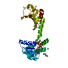
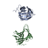
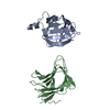
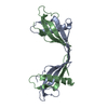
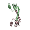
| Title | Crystal structure of the pleckstrin homology domain of Saccharomyces cerevisiae Avo1, a TORC2 subunit, in the P3121 crystal form | ||||||
 Components Components | Target of rapamycin complex 2 subunit AVO1 | ||||||
 Keywords Keywords | MEMBRANE PROTEIN / PH domain / membrane localization | ||||||
| Function / homology |  Function and homology information Function and homology informationPIP3 activates AKT signaling / CD28 dependent PI3K/Akt signaling / High laminar flow shear stress activates signaling by PIEZO1 and PECAM1:CDH5:KDR in endothelial cells / Regulation of TP53 Degradation / establishment or maintenance of actin cytoskeleton polarity / VEGFR2 mediated vascular permeability / TORC2 complex / TORC2 signaling / vacuolar membrane / TOR signaling ...PIP3 activates AKT signaling / CD28 dependent PI3K/Akt signaling / High laminar flow shear stress activates signaling by PIEZO1 and PECAM1:CDH5:KDR in endothelial cells / Regulation of TP53 Degradation / establishment or maintenance of actin cytoskeleton polarity / VEGFR2 mediated vascular permeability / TORC2 complex / TORC2 signaling / vacuolar membrane / TOR signaling / phosphatidylinositol-4,5-bisphosphate binding / regulation of cell growth / molecular adaptor activity / plasma membrane / cytoplasm Similarity search - Function | ||||||
| Biological species |  | ||||||
| Method |  X-RAY DIFFRACTION / SYNCHROTRON / MOLECULAR REPLACEMENT / Resolution: 2.8 Å X-RAY DIFFRACTION / SYNCHROTRON / MOLECULAR REPLACEMENT / Resolution: 2.8 Å | ||||||
 Authors Authors | Pan, D. / Matsuura, Y. | ||||||
 Citation Citation | Journal: Acta Crystallogr.,Sect.F / Year: 2012 Title: Structures of the pleckstrin homology domain of Saccharomyces cerevisiae Avo1 and its human orthologue Sin1, an essential subunit of TOR complex 2 Authors: Pan, D. / Matsuura, Y. | ||||||
| History |
|
- Structure visualization
| Structure viewer | Molecule: MolmilJmol/JSmol |
|---|
- Downloads & links
-Download
| PDBx/mmCIF format | 3ulc.cif.gz | 33.8 KB | Display | PDBx/mmCIF format |
|---|---|---|---|---|
| PDB format | pdb3ulc.ent.gz | 21.7 KB | Display | PDB format |
| PDBx/mmJSON format | 3ulc.json.gz | Tree view | PDBx/mmJSON format | |
| Others |  Other downloads Other downloads |
-Validation report
| Arichive directory | https://data.pdbj.org/pub/pdb/validation_reports/ul/3ulcftp://data.pdbj.org/pub/pdb/validation_reports/ul/3ulc | HTTPS FTP |
|---|
-Related structure data
| Related structure data |  3ulbSC 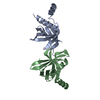 3voqC S: Starting model for refinement C: citing same article ( |
|---|---|
| Similar structure data |

-Links
 PDBj
PDBj

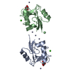
- Assembly
| Deposited unit | 
| ||||||||
|---|---|---|---|---|---|---|---|---|---|
| 1 | 
| ||||||||
| Unit cell |
|
-Components
| #1: Protein | Mass: 14608.734 Da / Num. of mol.: 1 / Fragment: PH domain, residues 1056-1176 Source method: isolated from a genetically manipulated source Source: (gene. exp.) Strain: S288c / Gene: AVO1 / Production host:  |
|---|---|
| #2: Water | ChemComp-HOH /  Mass: 18.015 Da / Num. of mol.: 2 / Source method: isolated from a natural source / Formula: H2O Mass: 18.015 Da / Num. of mol.: 2 / Source method: isolated from a natural source / Formula: H2O |
-Experimental details
-Experiment
| Experiment | Method: X-RAY DIFFRACTION / Number of used crystals: 1 |
|---|
- Sample preparation
| Crystal | Density Matthews: 3.08 Å3/Da / Density % sol: 60.11 % |
|---|---|
| Crystal grow | Temperature: 293 K / Method: vapor diffusion, sitting drop / pH: 7 Details: 0.1M Tris-HCl, 6% PEG8000, pH 7.0, VAPOR DIFFUSION, SITTING DROP, temperature 293K |
-Data collection
| Diffraction | Mean temperature: 100 K |
|---|---|
| Diffraction source | Source: SYNCHROTRON / Site: Photon Factory  / Beamline: BL-5A / Wavelength: 1 Å / Beamline: BL-5A / Wavelength: 1 Å |
| Detector | Type: ADSC QUANTUM 315r / Detector: CCD / Date: Oct 20, 2009 |
| Radiation | Monochromator: Numerical link type Si(111) double crystal monochromator Protocol: SINGLE WAVELENGTH / Monochromatic (M) / Laue (L): M / Scattering type: x-ray |
| Radiation wavelength | Wavelength: 1 Å / Relative weight: 1 |
| Reflection | Resolution: 2.8→32.88 Å / Num. all: 4709 / Num. obs: 4563 / % possible obs: 96.9 % / Observed criterion σ(F): 0 / Observed criterion σ(I): 0 / Redundancy: 9.8 % / Rmerge(I) obs: 0.051 |
| Reflection shell | Resolution: 2.8→2.95 Å / Redundancy: 8.2 % / Rmerge(I) obs: 0.595 / Mean I/σ(I) obs: 3.7 / % possible all: 84 |
- Processing
| Software |
| |||||||||||||||||||||||||||||||||||||||||||||
|---|---|---|---|---|---|---|---|---|---|---|---|---|---|---|---|---|---|---|---|---|---|---|---|---|---|---|---|---|---|---|---|---|---|---|---|---|---|---|---|---|---|---|---|---|---|---|
| Refinement | Method to determine structure: MOLECULAR REPLACEMENT Starting model: PDB entry 3ULB Resolution: 2.8→28.64 Å / Cor.coef. Fo:Fc: 0.907 / Cor.coef. Fo:Fc free: 0.838 / Occupancy max: 1 / Occupancy min: 1 / SU B: 15 / SU ML: 0.281 / Cross valid method: THROUGHOUT / σ(F): 0 / ESU R Free: 0.358 / Stereochemistry target values: MAXIMUM LIKELIHOOD Details: HYDROGENS HAVE BEEN USED IF PRESENT IN THE INPUT U VALUES: REFINED INDIVIDUALLY
| |||||||||||||||||||||||||||||||||||||||||||||
| Solvent computation | Ion probe radii: 0.8 Å / Shrinkage radii: 0.8 Å / VDW probe radii: 1.2 Å / Solvent model: MASK | |||||||||||||||||||||||||||||||||||||||||||||
| Displacement parameters | Biso max: 123.71 Å2 / Biso mean: 57.7293 Å2 / Biso min: 24.63 Å2
| |||||||||||||||||||||||||||||||||||||||||||||
| Refinement step | Cycle: LAST / Resolution: 2.8→28.64 Å
| |||||||||||||||||||||||||||||||||||||||||||||
| Refine LS restraints |
| |||||||||||||||||||||||||||||||||||||||||||||
| LS refinement shell | Resolution: 2.8→2.873 Å / Total num. of bins used: 20
|
