 Movie
Movie Controller

[English] 日本語
 Yorodumi
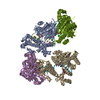
- PDB-3udu: Crystal structure of putative 3-isopropylmalate dehydrogenase fro... -
+ Open data

- Basic information
| Entry | Database: PDB / ID: 3udu | ||||||
|---|---|---|---|---|---|---|---|
| Title | Crystal structure of putative 3-isopropylmalate dehydrogenase from Campylobacter jejuni | ||||||
 Components Components | 3-isopropylmalate dehydrogenase | ||||||
 Keywords Keywords | OXIDOREDUCTASE / Structural Genomics / Center for Structural Genomics of Infectious Diseases / CSGID / putative isopropylmalate dehydrogenase | ||||||
| Function / homology |  Function and homology information Function and homology information3-isopropylmalate dehydrogenase / 3-isopropylmalate dehydrogenase activity / L-leucine biosynthetic process / NAD binding / magnesium ion binding / cytosol Similarity search - Function | ||||||
| Biological species |   Campylobacter jejuni (Campylobacter) Campylobacter jejuni (Campylobacter) | ||||||
| Method |  X-RAY DIFFRACTION / SYNCHROTRON / MIR / Resolution: 1.85 Å X-RAY DIFFRACTION / SYNCHROTRON / MIR / Resolution: 1.85 Å | ||||||
 Authors Authors | Tkaczuk, K.L. / Chruszcz, M. / Grimshaw, S. / Onopriyenko, O. / Savchenko, A. / Anderson, W.F. / Minor, W. / Center for Structural Genomics of Infectious Diseases (CSGID) | ||||||
 Citation Citation | Journal: To be Published Title: Crystal structure of putative 3-isopropylmalate dehydrogenase from Campylobacter jejuni Authors: Tkaczuk, K.L. / Chruszcz, M. / Grimshaw, S. / Onopriyenko, O. / Savchenko, A. / Anderson, W.F. / Minor, W. / Center for Structural Genomics of Infectious Diseases (CSGID) | ||||||
| History |
|
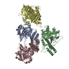
- Structure visualization
| Structure viewer | Molecule: MolmilJmol/JSmol |
|---|
- Downloads & links
-Download
| PDBx/mmCIF format | 3udu.cif.gz | 572.5 KB | Display | PDBx/mmCIF format |
|---|---|---|---|---|
| PDB format | pdb3udu.ent.gz | 468.7 KB | Display | PDB format |
| PDBx/mmJSON format | 3udu.json.gz | Tree view | PDBx/mmJSON format | |
| Others |  Other downloads Other downloads |
-Validation report
| Arichive directory | https://data.pdbj.org/pub/pdb/validation_reports/ud/3uduftp://data.pdbj.org/pub/pdb/validation_reports/ud/3udu | HTTPS FTP |
|---|
-Related structure data
| Related structure data | 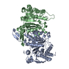 1cm7S S: Starting model for refinement |
|---|---|
| Similar structure data | |
| Other databases |

-Links
 PDBj
PDBj

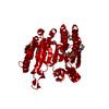
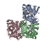
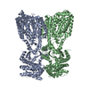
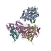
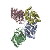
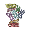
- Assembly
| Deposited unit | 
| ||||||||
|---|---|---|---|---|---|---|---|---|---|
| 1 | 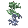
| ||||||||
| 2 | 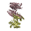
| ||||||||
| 3 | 
| ||||||||
| 4 | 
| ||||||||
| Unit cell |
|
-Components
| #1: Protein | Mass: 39649.438 Da / Num. of mol.: 8 Source method: isolated from a genetically manipulated source Source: (gene. exp.) Campylobacter jejuni (Campylobacter) / Gene: leuB, Cj1718c / Plasmid: pMCSG7 / Production host: References: UniProt: Q9PLW0, 3-isopropylmalate dehydrogenase #2: Chemical | ChemComp-EDO /   Mass: 62.068 Da / Num. of mol.: 6 / Source method: obtained synthetically / Formula: C2H6O2 Mass: 62.068 Da / Num. of mol.: 6 / Source method: obtained synthetically / Formula: C2H6O2#3: Chemical | ChemComp-CL / |   Mass: 35.453 Da / Num. of mol.: 1 / Source method: obtained synthetically / Formula: Cl Mass: 35.453 Da / Num. of mol.: 1 / Source method: obtained synthetically / Formula: Cl#4: Water | ChemComp-HOH / |  Mass: 18.015 Da / Num. of mol.: 1816 / Source method: isolated from a natural source / Formula: H2O Mass: 18.015 Da / Num. of mol.: 1816 / Source method: isolated from a natural source / Formula: H2O |
|---|
-Experimental details
-Experiment
| Experiment | Method: X-RAY DIFFRACTION / Number of used crystals: 1 |
|---|
- Sample preparation
| Crystal | Density Matthews: 2.17 Å3/Da / Density % sol: 43.39 % |
|---|---|
| Crystal grow | Temperature: 297 K / Method: vapor diffusion, hanging drop / pH: 7 Details: 25%PEG3350, 0.2M Na Chloride, 0.1MHEPES, pH 7, VAPOR DIFFUSION, HANGING DROP, temperature 297K |
-Data collection
| Diffraction | Mean temperature: 100 K |
|---|---|
| Diffraction source | Source: SYNCHROTRON / Site: APS  / Beamline: 19-ID / Wavelength: 0.9793 Å / Beamline: 19-ID / Wavelength: 0.9793 Å |
| Detector | Type: ADSC QUANTUM 315r / Detector: CCD / Date: Mar 19, 2010 |
| Radiation | Monochromator: Si 111 channel / Protocol: SINGLE WAVELENGTH / Monochromatic (M) / Laue (L): M / Scattering type: x-ray |
| Radiation wavelength | Wavelength: 0.9793 Å / Relative weight: 1 |
| Reflection | Resolution: 1.85→50 Å / Num. all: 213853 / Num. obs: 213853 / % possible obs: 99.7 % / Observed criterion σ(F): 0 / Observed criterion σ(I): -3 / Redundancy: 2.5 % / Biso Wilson estimate: 30.7 Å2 / Rmerge(I) obs: 0.093 / Rsym value: 0.093 / Net I/σ(I): 18.2 |
| Reflection shell | Resolution: 1.85→1.88 Å / Redundancy: 2.5 % / Rmerge(I) obs: 0.554 / Mean I/σ(I) obs: 2 / Num. unique all: 10781 / Rsym value: 0.554 / % possible all: 94.7 |
- Processing
| Software |
| ||||||||||||||||||||||||||||||||||||||||||||||||||||||||||||
|---|---|---|---|---|---|---|---|---|---|---|---|---|---|---|---|---|---|---|---|---|---|---|---|---|---|---|---|---|---|---|---|---|---|---|---|---|---|---|---|---|---|---|---|---|---|---|---|---|---|---|---|---|---|---|---|---|---|---|---|---|---|
| Refinement | Method to determine structure: MIR Starting model: PDB ENTRY 1CM7 Resolution: 1.85→50 Å / Cor.coef. Fo:Fc: 0.962 / Cor.coef. Fo:Fc free: 0.946 / Cross valid method: THROUGHOUT / σ(F): 0 / σ(I): 0 / ESU R Free: 0.154 / Stereochemistry target values: MAXIMUM LIKELIHOOD / Details: HYDROGENS HAVE BEEN ADDED IN THE RIDING POSITIONS
| ||||||||||||||||||||||||||||||||||||||||||||||||||||||||||||
| Solvent computation | Ion probe radii: 0.8 Å / Shrinkage radii: 0.8 Å / VDW probe radii: 1.2 Å / Solvent model: BABINET MODEL WITH MASK | ||||||||||||||||||||||||||||||||||||||||||||||||||||||||||||
| Displacement parameters | Biso mean: 24.991 Å2
| ||||||||||||||||||||||||||||||||||||||||||||||||||||||||||||
| Refinement step | Cycle: LAST / Resolution: 1.85→50 Å
| ||||||||||||||||||||||||||||||||||||||||||||||||||||||||||||
| Refine LS restraints |
| ||||||||||||||||||||||||||||||||||||||||||||||||||||||||||||
| LS refinement shell | Resolution: 1.85→1.898 Å / Total num. of bins used: 20
|
