 Movie
Movie Controller

[English] 日本語
 Yorodumi
- PDB-5khn: Crystal structures of the Burkholderia multivorans hopanoid trans... -
+ Open data

- Basic information
| Entry | Database: PDB / ID: 5khn | ||||||
|---|---|---|---|---|---|---|---|
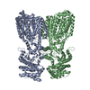
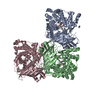
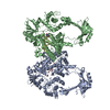
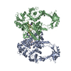
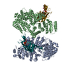
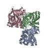
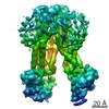
| Title | Crystal structures of the Burkholderia multivorans hopanoid transporter HpnN | ||||||
 Components Components | RND transporter | ||||||
 Keywords Keywords | MEMBRANE PROTEIN / transmembrane helices | ||||||
| Function / homology | :  Function and homology information Function and homology information | ||||||
| Biological species |  Burkholderia multivorans (bacteria) Burkholderia multivorans (bacteria) | ||||||
| Method |  X-RAY DIFFRACTION / SYNCHROTRON / SIRAS / Resolution: 3.445 Å X-RAY DIFFRACTION / SYNCHROTRON / SIRAS / Resolution: 3.445 Å | ||||||
 Authors Authors | Su, C.-C. / Yu, E.W. | ||||||
 Citation Citation | Journal: Proc. Natl. Acad. Sci. U.S.A. / Year: 2017 Title: Crystal structures of the Burkholderia multivorans hopanoid transporter HpnN. Authors: Kumar, N. / Su, C.C. / Chou, T.H. / Radhakrishnan, A. / Delmar, J.A. / Rajashankar, K.R. / Yu, E.W. | ||||||
| History |
|
- Structure visualization
| Structure viewer | Molecule: MolmilJmol/JSmol |
|---|
- Downloads & links
-Download
| PDBx/mmCIF format | 5khn.cif.gz | 316.1 KB | Display | PDBx/mmCIF format |
|---|---|---|---|---|
| PDB format | pdb5khn.ent.gz | 256.7 KB | Display | PDB format |
| PDBx/mmJSON format | 5khn.json.gz | Tree view | PDBx/mmJSON format | |
| Others |  Other downloads Other downloads |
-Validation report
| Arichive directory | https://data.pdbj.org/pub/pdb/validation_reports/kh/5khnftp://data.pdbj.org/pub/pdb/validation_reports/kh/5khn | HTTPS FTP |
|---|
-Related structure data


-Links
 PDBj
PDBj- Assembly
| Deposited unit | 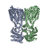
| ||||||||
|---|---|---|---|---|---|---|---|---|---|
| 1 |
| ||||||||
| Unit cell |
|
-Components
| #1: Protein | Mass: 93501.477 Da / Num. of mol.: 2 / Mutation: R362A Source method: isolated from a genetically manipulated source Source: (gene. exp.) Burkholderia multivorans (bacteria) / Gene: WM33_10510 / Plasmid: pET15b / Production host: |
|---|
-Experimental details
-Experiment
| Experiment | Method: X-RAY DIFFRACTION / Number of used crystals: 1 |
|---|
- Sample preparation
| Crystal | Density Matthews: 4 Å3/Da / Density % sol: 69.25 % |
|---|---|
| Crystal grow | Temperature: 298 K / Method: vapor diffusion / pH: 3.5 / Details: 18% PEG2000, and 0.2M LiSo4 |
-Data collection
| Diffraction | Mean temperature: 100 K | |||||||||||||||||||||||||||||||||||||||||||||||||||||||||||||||||||||||||||||||||||||||||||||||||||
|---|---|---|---|---|---|---|---|---|---|---|---|---|---|---|---|---|---|---|---|---|---|---|---|---|---|---|---|---|---|---|---|---|---|---|---|---|---|---|---|---|---|---|---|---|---|---|---|---|---|---|---|---|---|---|---|---|---|---|---|---|---|---|---|---|---|---|---|---|---|---|---|---|---|---|---|---|---|---|---|---|---|---|---|---|---|---|---|---|---|---|---|---|---|---|---|---|---|---|---|---|
| Diffraction source | Source: SYNCHROTRON / Site: APS  / Beamline: 24-ID-C / Wavelength: 0.98 Å / Beamline: 24-ID-C / Wavelength: 0.98 Å | |||||||||||||||||||||||||||||||||||||||||||||||||||||||||||||||||||||||||||||||||||||||||||||||||||
| Detector | Type: DECTRIS PILATUS3 S 6M / Detector: PIXEL / Date: Feb 27, 2016 | |||||||||||||||||||||||||||||||||||||||||||||||||||||||||||||||||||||||||||||||||||||||||||||||||||
| Radiation | Monochromator: Si(III) / Protocol: SINGLE WAVELENGTH / Monochromatic (M) / Laue (L): M / Scattering type: x-ray | |||||||||||||||||||||||||||||||||||||||||||||||||||||||||||||||||||||||||||||||||||||||||||||||||||
| Radiation wavelength | Wavelength: 0.98 Å / Relative weight: 1 | |||||||||||||||||||||||||||||||||||||||||||||||||||||||||||||||||||||||||||||||||||||||||||||||||||
| Reflection | Resolution: 3.45→100 Å / Num. obs: 38588 / % possible obs: 99.9 % / Redundancy: 11 % / Biso Wilson estimate: 126.04 Å2 / Rmerge(I) obs: 0.126 / Rpim(I) all: 0.051 / Rrim(I) all: 0.132 / Χ2: 1.227 / Net I/σ(I): 7.3 / Num. measured all: 424764 | |||||||||||||||||||||||||||||||||||||||||||||||||||||||||||||||||||||||||||||||||||||||||||||||||||
| Reflection shell | Diffraction-ID: 1
|
- Processing
| Software |
| |||||||||||||||||||||||||||||||||||||||||||||||||||||||||||||||||||||||||||||||||||||||||||||||||||||||||
|---|---|---|---|---|---|---|---|---|---|---|---|---|---|---|---|---|---|---|---|---|---|---|---|---|---|---|---|---|---|---|---|---|---|---|---|---|---|---|---|---|---|---|---|---|---|---|---|---|---|---|---|---|---|---|---|---|---|---|---|---|---|---|---|---|---|---|---|---|---|---|---|---|---|---|---|---|---|---|---|---|---|---|---|---|---|---|---|---|---|---|---|---|---|---|---|---|---|---|---|---|---|---|---|---|---|---|
| Refinement | Method to determine structure: SIRAS / Resolution: 3.445→93.803 Å / SU ML: 0.72 / Cross valid method: FREE R-VALUE / σ(F): 1.34 / Phase error: 40.31 / Stereochemistry target values: ML
| |||||||||||||||||||||||||||||||||||||||||||||||||||||||||||||||||||||||||||||||||||||||||||||||||||||||||
| Solvent computation | Shrinkage radii: 0.9 Å / VDW probe radii: 1.11 Å / Solvent model: FLAT BULK SOLVENT MODEL | |||||||||||||||||||||||||||||||||||||||||||||||||||||||||||||||||||||||||||||||||||||||||||||||||||||||||
| Displacement parameters | Biso max: 217.92 Å2 / Biso mean: 127.5209 Å2 / Biso min: 30 Å2 | |||||||||||||||||||||||||||||||||||||||||||||||||||||||||||||||||||||||||||||||||||||||||||||||||||||||||
| Refinement step | Cycle: final / Resolution: 3.445→93.803 Å
| |||||||||||||||||||||||||||||||||||||||||||||||||||||||||||||||||||||||||||||||||||||||||||||||||||||||||
| Refine LS restraints |
| |||||||||||||||||||||||||||||||||||||||||||||||||||||||||||||||||||||||||||||||||||||||||||||||||||||||||
| LS refinement shell | Refine-ID: X-RAY DIFFRACTION / Rfactor Rfree error: 0 / Total num. of bins used: 14
|
